Visual acuity is a measure of:
Light entering the eye passes through which retinal layer first?
Synchysis refers to
A 52-year-old man presents with painless, gradual vision loss in his right eye over two hours. He has a history of myocardial infarction and takes aspirin daily. The physical examination reveals no vision in the right eye. At the molecular level, which of the following components is essential for the first step of the visual cascade?
Which is the longest and thinnest extraocular muscle?
All statements are true about the eye of a newborn except?
In near vision, what change occurs in the eye?
A person with defective blue color appreciation is called?
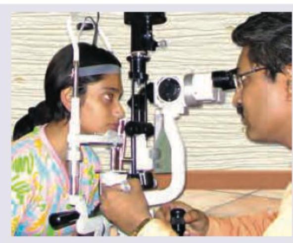
All of the following are correct about the image shown except: (Recent NEET Pattern 2016-17)

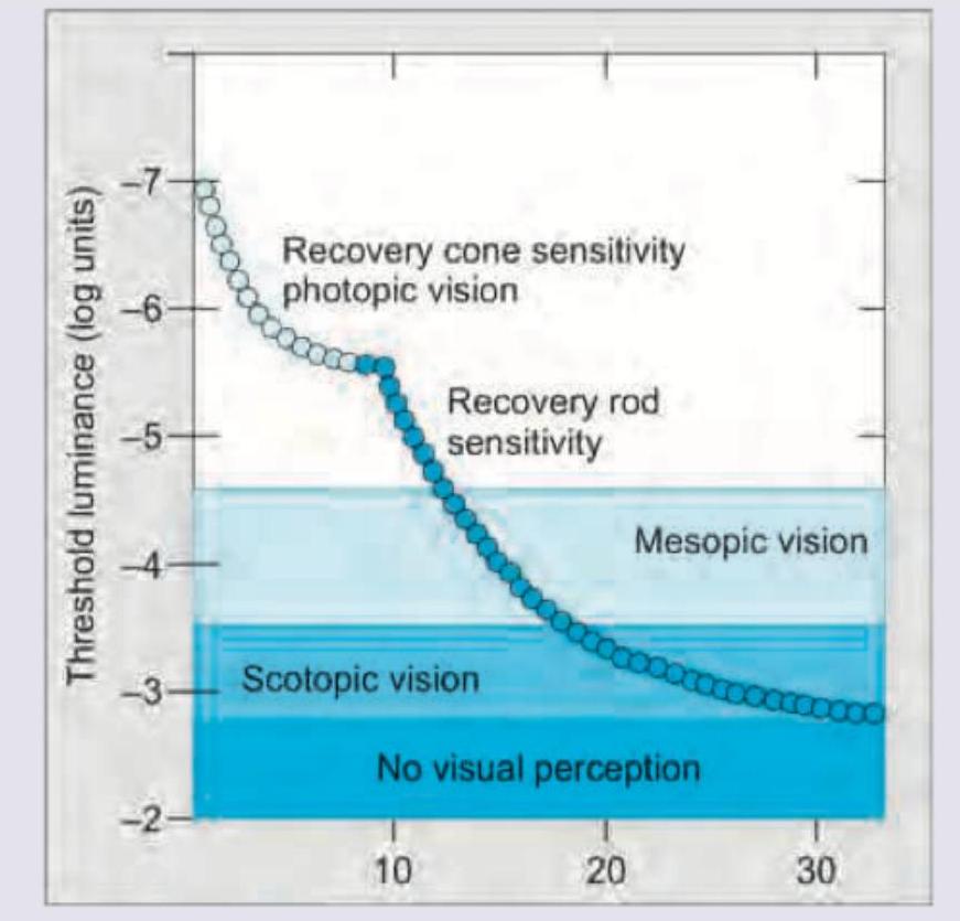
The image given below shows:

Explanation: **Explanation:** Visual acuity is defined as the ability of the eye to distinguish between two separate points in space, which is the fundamental basis of **Form Sense**. **1. Why "Form Sense" is Correct:** Form sense is the faculty of the eye to perceive the shape and contour of objects. It is a function of the **fovea centralis**, where the density of cones is highest. Visual acuity (tested via Snellen’s chart) specifically measures the **minimum angle of resolution (MAR)**—the smallest angle at which two points can be perceived as distinct. For a person with 6/6 vision, this angle is 1 minute of arc. **2. Why other options are incorrect:** * **Light Sense (Option A):** This refers to the ability to perceive light and differentiate between different intensities of illumination. It is primarily a function of the rods and is tested via dark adaptation and light threshold tests. * **Contrast Sense (Option C):** This is the ability to distinguish an object from its background. While related to visual quality, it is a separate parameter measured using Pelli-Robson or CSV-1000 charts. It is often affected earlier than visual acuity in conditions like glaucoma or optic neuritis. **Clinical Pearls for NEET-PG:** * **Minimum Cognizable:** The ability to identify a shape (e.g., a letter on a Snellen chart). * **Minimum Separable:** The ability to see two points as separate (the basis of the Landolt C test). * **Anatomical Basis:** Form sense is best developed at the fovea; Light sense is best in the periphery (rods). * **Visual Angle:** The standard Snellen letter subtends an angle of 5 minutes at the nodal point, while each individual arm/gap subtends 1 minute.
Explanation: **Explanation:** To understand the path of light through the retina, one must recognize that the human retina is **"inverted."** Although the photoreceptors (rods and cones) are the cells that actually detect light, they are located in the outermost layer, adjacent to the Retinal Pigment Epithelium (RPE). **Why the Retinal Ganglion Layer is correct:** When light enters the eye and hits the retina, it must travel through almost the entire thickness of the sensory retina before reaching the photoreceptors. The **Retinal Ganglion Cell (RGC) layer** is the innermost cellular layer (closest to the vitreous). Therefore, light passes through the Internal Limiting Membrane and the Nerve Fiber Layer first, followed immediately by the **Ganglion Cell Layer**, before reaching the deeper nuclear and plexiform layers. **Analysis of Incorrect Options:** * **Inner Nuclear Layer (A):** This contains the cell bodies of bipolar, horizontal, and amacrine cells. It is located deeper than the ganglion cell layer. * **Outer Plexiform Layer (C):** This is the area of synapse between photoreceptors and bipolar/horizontal cells. Light reaches this only after passing the inner layers. * **Outer Nuclear Layer (B):** This contains the cell bodies of the rods and cones. It is one of the last layers light reaches before hitting the photoreceptor outer segments. **High-Yield NEET-PG Pearls:** * **Path of Light:** Vitreous → Nerve Fiber Layer → Ganglion Cell Layer → ... → Photoreceptors → RPE. * **Path of Nerve Impulse:** Photoreceptors → Bipolar Cells → Ganglion Cells → Optic Nerve (Reverse of the light path). * **Foveola Fact:** At the foveola (the center of the macula), the inner retinal layers are displaced laterally. This allows light to strike the photoreceptors directly, minimizing scattering and providing the highest visual acuity.
Explanation: **Explanation:** **Synchysis** is the medical term for the **liquefaction of the vitreous humor**. The vitreous is a transparent, gel-like structure composed primarily of water (99%), collagen fibrils, and hyaluronic acid. As the eye ages or due to certain pathological conditions, the collagen framework collapses, and the hyaluronic acid releases its bound water, resulting in the formation of liquid pockets within the gel. * **Why Option C is correct:** Synchysis specifically describes this biochemical and structural transition from a gel state to a liquid state. It is most commonly seen as a senile change (age-related) or in pathological myopia. * **Why Option A is wrong:** Collapse of the vitreous is termed **Syneresis**. While synchysis (liquefaction) often leads to syneresis, they are distinct processes. * **Why Option B is wrong:** Black spots in front of the eyes are known as **Muscae volitantes** (floaters). These are the clinical symptoms caused by opacities casting shadows on the retina, often resulting from synchysis. * **Why Option D is wrong:** Detachment of the vitreous from the internal limiting membrane of the retina is called **Posterior Vitreous Detachment (PVD)**. **High-Yield Clinical Pearls for NEET-PG:** 1. **Synchysis Scintillans:** A condition where cholesterol crystals are found in a liquefied vitreous (usually post-traumatic or post-inflammatory). These crystals settle at the bottom due to gravity but shower upward like a "snow globe" when the eye moves. 2. **Asteroid Hyalosis:** Characterized by calcium-lipid complexes (soaps) suspended in a *solid* vitreous. Unlike synchysis scintillans, these do not settle. 3. **Wagner’s Disease:** A genetic condition characterized by "optically empty vitreous" due to extreme synchysis.
Explanation: ### Explanation The visual cycle (phototransduction) begins in the outer segments of photoreceptors (rods and cones). The fundamental molecular event that triggers the visual cascade is the **photoisomerization** of the chromophore. **1. Why 11-cis-retinal is correct:** In the dark, the visual pigment **Rhodopsin** consists of a protein (opsin) bound to **11-cis-retinal**, which acts as an inverse agonist, keeping the receptor inactive. When a photon of light strikes the retina, it is absorbed by 11-cis-retinal, causing it to undergo an instantaneous conformational change into **all-trans-retinal**. This is the **first and only light-dependent step** in the entire visual process. **2. Why the other options are incorrect:** * **B. All-cis-retinal:** This is a non-physiological isomer and does not play a role in the human visual cycle. * **C. All-trans-retinal:** This is the *product* of the first step, not the starting component. After isomerization, it must be transported to the Retinal Pigment Epithelium (RPE) to be recycled back into 11-cis-retinal. * **D. Meta-rhodopsin II:** Also known as "activated rhodopsin," this is an intermediate biochemical state formed *after* the isomerization of retinal. It is responsible for activating the G-protein (Transducin), but it is not the initial component. **Clinical Pearls & High-Yield Facts for NEET-PG:** * **Vitamin A (Retinol):** The precursor to 11-cis-retinal. Deficiency leads to Nyctalopia (night blindness) because rods are affected first. * **RPE65:** The key enzyme in the RPE that converts all-trans-retinyl ester to 11-cis-retinol. Mutations in this enzyme are associated with Leber Congenital Amaurosis. * **Clinical Correlation:** While the question describes a clinical scenario suggestive of **Central Retinal Artery Occlusion (CRAO)** (painless sudden loss of vision in an elderly patient with cardiovascular risk), the molecular question focuses on the physiology of vision. In CRAO, the "cherry-red spot" occurs because the fovea receives blood from the ciliary circulation, while the surrounding retina becomes pale due to edema.
Explanation: **Explanation:** The **Superior Oblique (SO)** is the correct answer because it is anatomically the longest and thinnest extraocular muscle. It measures approximately **58 mm to 60 mm** in total length (32 mm for the muscular portion and 26 mm to 28 mm for the tendon). Its thin, cord-like tendon passes through the **trochlea** (a fibrocartilaginous pulley), which allows it to change direction and insert into the posterolateral aspect of the globe. **Analysis of Options:** * **Superior Rectus (SR):** While it is the longest of the *recti* muscles (approx. 40 mm), it is significantly shorter than the Superior Oblique. * **Inferior Rectus (IR):** This is generally considered the shortest of the recti muscles (approx. 40 mm) and is thicker than the SO. * **Inferior Oblique (IO):** This is the shortest extraocular muscle overall (approx. 37 mm) and is the only muscle that does not originate from the Apex of the orbit (it originates from the orbital floor). **High-Yield Clinical Pearls for NEET-PG:** * **Innervation:** The SO is the only muscle supplied by the **Trochlear nerve (CN IV)**. It is also the only extraocular muscle whose nerve enters from the orbital (outer) surface rather than the internal surface. * **Action:** The primary action of the SO is **Intorsion** (remember: "Superior muscles are Intorters"). Its secondary actions are depression and abduction. * **Origin:** Like the recti, it originates from the Annulus of Zinn, but it is the only muscle to pass through a pulley (Trochlea). * **Shortest Muscle:** Always remember the **Inferior Oblique** is the shortest, while the **Superior Oblique** is the longest.
Explanation: **Explanation:** The correct answer is **D**. A newborn is typically **hypermetropic** (farsighted), not myopic. The average refractive error at birth is approximately **+2.0 to +3.0 Diopters**. This occurs because the axial length of the newborn eye is short (approx. 17 mm), and the refractive power of the lens and cornea cannot fully compensate for this shortness. As the child grows, the eye undergoes "emmetropization." **Analysis of other options:** * **Option A (True):** Myelination of the optic nerve starts at the LGN and proceeds towards the eye. It reaches the **lamina cribrosa** at birth and normally stops there. If it continues into the nerve fiber layer, it results in "opaque/myelinated nerve fibers." * **Option B (True):** In newborns, the orbital axes are more divergent (approx. **50°**) compared to adults (approx. **45°**). This contributes to the appearance of pseudo-exotropia in some infants. * **Option C (True):** The retina is anatomically developed at birth except for the **macula**. The fovea centralis does not fully mature until about 4–6 months after birth, which is why central fixation is not present at birth. **High-Yield Clinical Pearls for NEET-PG:** * **Axial Length:** ~17 mm at birth; reaches adult size (~24 mm) by age 13–15. * **Corneal Diameter:** ~9.5–10 mm at birth. If >11 mm, suspect infantile glaucoma (Buphthalmos). It reaches the adult size of 11.7 mm by age 2. * **Lens:** More spherical at birth; has a higher refractive power (~30D) compared to an adult (~18-20D). * **Anterior Chamber:** Shallower in newborns compared to adults.
Explanation: To understand the changes in the eye during near vision, we must look at the **Near Reflex triad**: Accommodation, Convergence, and Miosis. ### **Why the correct answer is right:** **B. The depth of focus increases:** During near vision, the pupils constrict (**Miosis**). A smaller pupil acts like a pinhole camera, reducing the size of the blur circles on the retina. This physical phenomenon increases the **depth of focus** (the range over which an object can be moved without appearing out of focus), allowing for sharper vision of near objects. ### **Why the other options are wrong:** * **A. The power of the lens decreases:** Incorrect. During accommodation, the ciliary muscle contracts, causing the lens to become more spherical (convex). This **increases** the refractive power of the lens to focus divergent rays from near objects onto the retina. * **C. The lateral recti contract:** Incorrect. To maintain single binocular vision at a near point, the eyes must turn inward. This requires the contraction of both **medial recti** (Convergence), not the lateral recti. * **D. The zonular tension increases:** Incorrect. When the ciliary muscle contracts for near vision, it moves forward and inward, which **relaxes the suspensory ligaments (zonules)**. This relaxation allows the elastic lens capsule to bulge and increase its curvature. ### **High-Yield Clinical Pearls for NEET-PG:** * **Accommodation Mechanism (Helmholtz Theory):** Ciliary muscle contraction → Zonular relaxation → Increased lens curvature (mostly anterior) → Increased dioptric power. * **Presbyopia:** A physiological loss of accommodation due to age-related decrease in lens elasticity and ciliary muscle efficiency. * **Argyll Robertson Pupil:** A classic neuro-ophthalmic condition where the **Light Reflex is lost but the Accommodation Reflex is present** (Prostitute's Pupil), often seen in neurosyphilis.
Explanation: ### Explanation The question tests the nomenclature of color vision deficiencies based on the **Trichromatic Theory**. Human color vision relies on three photopigments (cones): Erythrolabe (Red/Long-wave), Chlorolabe (Green/Medium-wave), and Cyanolabe (Blue/Short-wave). **Why Tritanomalous is correct:** The suffix **"-anomalous"** refers to a deficiency or "weakness" in a specific color pigment (trichromacy is preserved, but one pigment is altered). The prefix **"Trit-"** refers to the third pigment, which is **Blue**. Therefore, a person with **Tritanomalous** vision has a defective appreciation of blue light due to an altered spectral sensitivity of the blue cones. **Analysis of Incorrect Options:** * **Deuteranomalous (A):** This refers to a "green weakness." It is the most common type of color vision deficiency. * **Deuteranopia (B):** The suffix **"-anopia"** indicates a complete absence of a pigment. Deuteranopia is the total absence of green-sensitive cones ("green blindness"). * **Tritanopia (C):** This is the total absence of blue-sensitive cones ("blue blindness"). While it involves the blue spectrum, the question specifies "defective appreciation" (weakness) rather than total absence. **NEET-PG High-Yield Pearls:** 1. **Inheritance:** Most color blindness (Red-Green) is **X-linked recessive**. However, **Tritan** defects (Blue-Yellow) are rare and typically **Autosomal Dominant** (encoded on Chromosome 7). 2. **Kollner’s Rule:** * Outer retinal diseases/macular degeneration usually cause **Blue-Yellow** defects. * Optic nerve diseases (except Glaucoma) usually cause **Red-Green** defects. 3. **Screening:** **Ishihara Charts** are the most common screening tool but are primarily designed to detect Red-Green defects, not Blue-Yellow. 4. **Gold Standard:** The **Nagel Anomaloscope** is the definitive test to differentiate between dichromacy and anomalous trichromacy.
Explanation: ***Hruby lens of 30D for lens examination*** - A **Hruby lens is a -55D to -58.6D non-contact lens** used for **fundus examination**, not lens examination, and is definitely not 30D. - The image shows a **slit lamp biomicroscope**, which is used for examining the anterior and posterior segments of the eye with various contact and non-contact lenses, but the Hruby lens is specifically designed for **retinal/fundus viewing**. - This statement is **incorrect** on two counts: wrong diopter power and wrong purpose. *Uses direct and indirect illumination to view structures of anterior chamber* - The **slit lamp** indeed utilizes various illumination techniques, including **direct and indirect focal illumination**, to visualize different structures within the anterior chamber, such as the cornea, iris, and lens. - This versatility in illumination allows for detailed examination of both superficial and deeper ocular structures. - This statement is **correct**. *Used as delivery system for Argon and YAG laser* - The slit lamp serves as a common **delivery system for both Argon and Nd:YAG lasers** in ophthalmic procedures. - This allows for precise aiming and application of laser energy for conditions like **glaucoma (laser iridotomy, trabeculoplasty)** and **posterior capsulotomy**. - This statement is **correct**. *Accurate depth perception* - The **binocular viewing system** of the slit lamp provides excellent **stereopsis**, enabling the examiner to accurately assess the **depth and three-dimensional relationships** of ocular structures. - This feature is crucial for diagnosing conditions like **anterior chamber depth assessment, corneal foreign bodies, or cataract localization**. - This statement is **correct**.
Explanation: ***Dark adaptation curve*** - The image displays the change in **threshold luminance** over time, specifically showing two distinct phases of recovery of sensitivity: an initial rapid phase (cones) and a later, slower, more sensitive phase (rods). - This bimodal curve is characteristic of the **dark adaptation process**, where the eye adjusts from bright to dim light, increasing its sensitivity to light stimuli. *Visual evoked response* - A **visual evoked response (VER)** measures the electrical signals generated in the brain in response to visual stimuli. - VER graphs typically show amplitude and latency of brain activity, not a curve of threshold luminance over time. *Contrast sensitivity plot* - A **contrast sensitivity plot** illustrates the ability to distinguish between different levels of contrast at various spatial frequencies. - This is usually depicted as a curve showing contrast sensitivity as a function of spatial frequency, which is different from the time-dependent threshold luminance shown. *Electroretinography curve* - **Electroretinography (ERG)** measures the electrical responses of various retinal cell types to light stimuli. - An ERG curve typically shows a characteristic waveform with specific a-wave and b-wave components, representing photoreceptor and bipolar cell activity, which is not what is presented in the image.
Embryology of Eye
Practice Questions
Ocular Anatomy
Practice Questions
Ocular Physiology
Practice Questions
Ocular Biochemistry
Practice Questions
Ocular Microbiology
Practice Questions
Ocular Pharmacology
Practice Questions
Ocular Pathology
Practice Questions
Ocular Genetics
Practice Questions
Ocular Immunology
Practice Questions
Visual Neuroscience
Practice Questions
Ocular Imaging Physics
Practice Questions
Laser Physics in Ophthalmology
Practice Questions
Get full access to all questions, explanations, and performance tracking.
Start For Free