
Basic Sciences as Related to Eye — MCQs

On this page
Which statement is true about the central retinal artery?
Which is the thinnest part of the lens of the eye?
Light entering the eye passes through which retinal layer first?
Which of the following is NOT a function of aqueous humor?
All statements are true about the eye of a newborn except?
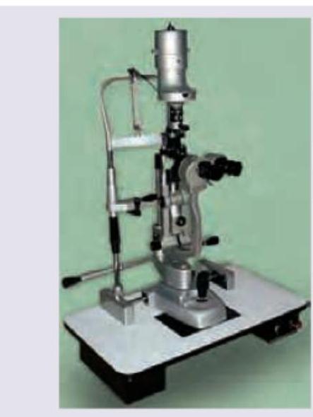
Which instrument is shown below?
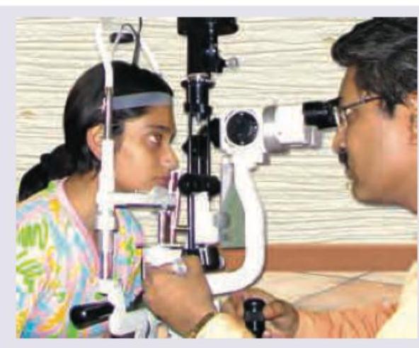
All of the following are correct about the image shown except: (Recent NEET Pattern 2016-17)
Pupil in acute iritis is:
Circumference of an adult eyeball is:
Father of ophthalmology?
Practice by Chapter
Embryology of Eye
Practice Questions
Ocular Anatomy
Practice Questions
Ocular Physiology
Practice Questions
Ocular Biochemistry
Practice Questions
Ocular Microbiology
Practice Questions
Ocular Pharmacology
Practice Questions
Ocular Pathology
Practice Questions
Ocular Genetics
Practice Questions
Ocular Immunology
Practice Questions
Visual Neuroscience
Practice Questions
Ocular Imaging Physics
Practice Questions
Laser Physics in Ophthalmology
Practice Questions
Want unlimited practice?
Get full access to all questions, explanations, and performance tracking.
Scan to download app