
Basic Sciences as Related to Eye — MCQs

On this page
A person is unable to look down. Which extraocular muscle is affected?
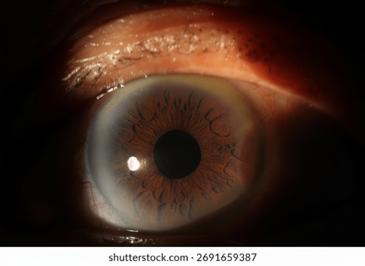
A 37-year-old woman presented with symptoms consistent with allergic conjunctivitis. What is the diagnosis for this incidental finding?
Which of the following is the most highly innervated structure of the eye?
Which of the following is contained within the posterior chamber of the eye?
The oily layer of the tear film is formed by which of the following?
Which atomic environment is used in the laser machines employed in ophthalmology?
Deep anterior chamber is seen in which of the following conditions?
Where is the circulus iridis major located?
What is the approximate normal volume of the anterior chamber of the eye?
Traumatic hyphaema (blood in the anterior chamber) occurs due to injury to the iris or ciliary body vessels. The circulus iridis major gives off branches to the ciliary processes. Which ocular muscle and nerve are primarily involved in controlling the medial rectus muscle?
Practice by Chapter
Embryology of Eye
Practice Questions
Ocular Anatomy
Practice Questions
Ocular Physiology
Practice Questions
Ocular Biochemistry
Practice Questions
Ocular Microbiology
Practice Questions
Ocular Pharmacology
Practice Questions
Ocular Pathology
Practice Questions
Ocular Genetics
Practice Questions
Ocular Immunology
Practice Questions
Visual Neuroscience
Practice Questions
Ocular Imaging Physics
Practice Questions
Laser Physics in Ophthalmology
Practice Questions
Want unlimited practice?
Get full access to all questions, explanations, and performance tracking.
Scan to download app