What is the best treatment for severe stress incontinence without prolapse?
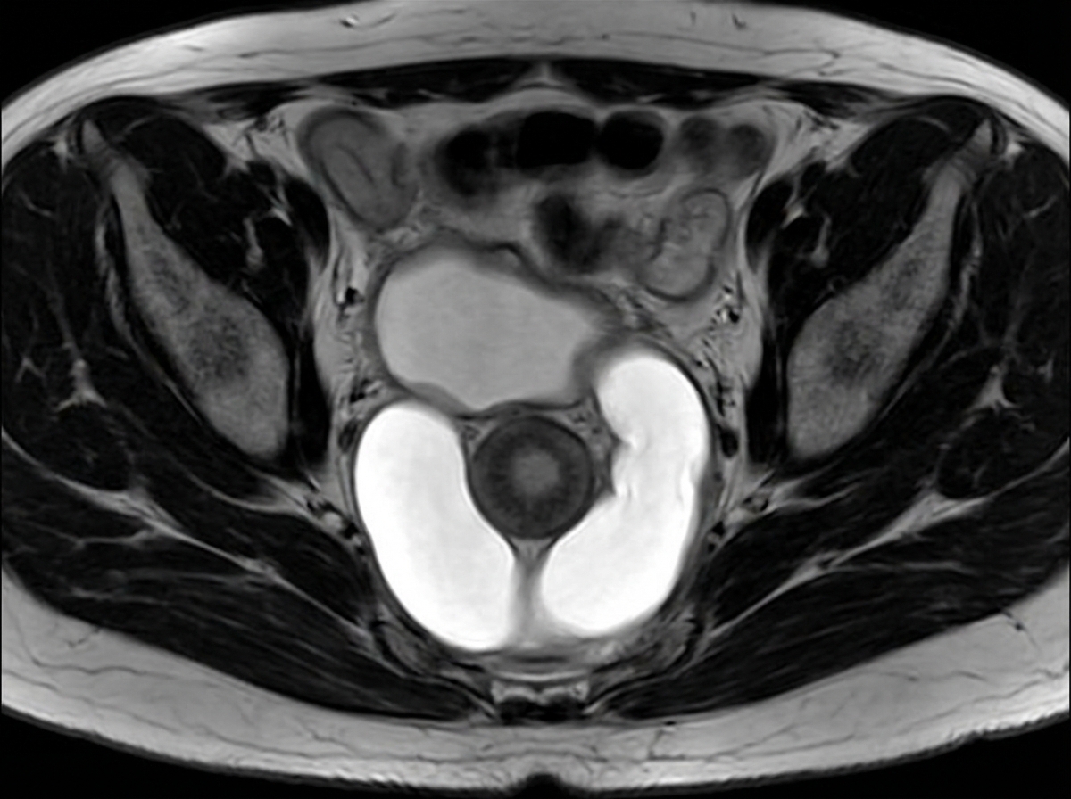
What is the treatment for genuine stress incontinence?
All of the following are vaginal procedures done for urinary incontinence, EXCEPT:
A 26-year-old nulliparous female presents with third-degree uterine prolapse, without associated cystocele or rectocele. What is the best treatment option?
Regarding bacterial vaginosis, all are true except?
What is the most useful investigation for vesicovaginal fistula (VVF)?
What is the most common type of urinary fistula?
A 65-year-old woman complains of leakage of urine. What is the most common cause of this condition in such patients?
Urinary incontinence in females is best investigated by:
Moschcowitz repair is done in case of?
Explanation: **Explanation:** The correct answer is **Tension-free vaginal tape (TVT)**. **Why it is correct:** Stress Urinary Incontinence (SUI) is primarily caused by urethral hypermobility or intrinsic sphincter deficiency. For **severe** SUI, surgical intervention is the gold standard. Mid-urethral slings (MUS), specifically **TVT**, have revolutionized treatment. The procedure involves placing a synthetic mesh under the mid-urethra to provide a "backstop" during increased intra-abdominal pressure (coughing/sneezing), mimicking the natural support of the pubourethral ligaments. It is currently considered the **gold standard** due to its high success rate (>90%), minimally invasive nature, and long-term efficacy. **Why other options are incorrect:** * **Pelvic floor exercises (Kegel’s):** These are the first-line treatment for *mild to moderate* SUI. However, for *severe* cases, they are often insufficient. * **Kelly’s repair:** This is an anterior colporrhaphy involving plication of the bladder neck. It is now considered **obsolete** for SUI because it has a high failure rate and is primarily used for correcting cystoceles, not incontinence. * **Burch colposuspension:** This was the previous gold standard. It involves anchoring the paravaginal fascia to Cooper’s ligament via an abdominal approach. While effective, it is more invasive than TVT and is now generally reserved for cases where abdominal surgery is already being performed for other reasons. **High-Yield Clinical Pearls for NEET-PG:** * **Investigation of choice:** Urodynamic study (to differentiate SUI from Urge Incontinence). * **Q-tip test:** A positive test (>30° angle change) indicates urethral hypermobility. * **TVT vs. TOT:** Transobturator Tape (TOT) is preferred in patients to avoid retropubic complications (like bladder perforation), but TVT is slightly superior for intrinsic sphincter deficiency. * **Gold Standard for SUI:** Mid-urethral slings (TVT/TOT).
Explanation: **Explanation:** **Genuine Stress Incontinence (GSI)** is defined as the involuntary leakage of urine when intra-abdominal pressure increases (e.g., coughing, sneezing) in the absence of detrusor contraction. The primary pathology is the loss of the posterior urethrovesical angle and hypermobility of the bladder neck. **Why Colposuspension is correct:** **Burch Colposuspension** is considered a gold-standard surgical treatment for GSI. It involves elevating the perivaginal fascia to the iliopectineal (Cooper’s) ligaments. This restores the bladder neck to an intra-abdominal position, allowing pressure transmission to the proximal urethra, thereby maintaining continence during physical exertion. **Analysis of Incorrect Options:** * **Anterior colporrhaphy (A):** This is the treatment for **cystocele**. While it was historically used for GSI (Kelly’s plication), it has high failure rates and is no longer the treatment of choice for incontinence. * **Posterior colporrhaphy (B):** This is the surgical repair for **rectocele** or deficient perineum; it has no role in treating urinary incontinence. * **Pelvic floor exercises (D):** Also known as Kegel exercises, these are the **first-line conservative management** for mild GSI. However, in the context of "treatment" (often implying definitive or surgical management in exam patterns when a gold-standard surgery is listed), Colposuspension is the superior surgical answer. **High-Yield Clinical Pearls for NEET-PG:** * **Gold Standard Surgery:** Mid-urethral slings (TVT - Tension-free Vaginal Tape or TOT - Transobturator Tape) are now more common than Burch Colposuspension due to being minimally invasive. * **Investigation of Choice:** Urodynamic study (Cystometry) is used to confirm GSI and rule out Urge Incontinence. * **Q-tip Test:** A positive test (>30-degree angle change) indicates urethral hypermobility. * **Marshall-Marchetti-Krantz (MMK):** An older procedure where the bladder neck is sutured to the periosteum of the pubic symphysis (now rarely done due to risk of osteitis pubis).
Explanation: **Explanation:** The core concept tested here is the **surgical approach** (Vaginal vs. Abdominal) used to treat Stress Urinary Incontinence (SUI). **1. Why Option C is Correct:** The **Marshall-Marchetti-Krantz (MMK) procedure** is a **retropubic abdominal surgery**. It involves suturing the periurethral tissues to the periosteum of the pubic symphysis. Because it requires an abdominal incision (Laparotomy), it is not a vaginal procedure. While effective, it has largely been replaced by the Burch colposuspension (which uses Cooper’s ligament) to avoid the risk of osteitis pubis. **2. Why the other options are Incorrect (Vaginal Procedures):** * **Transobturator Tape (TOT):** A modern "mid-urethral sling" procedure performed entirely through the **vaginal route** with small exit punctures in the groin. It is currently a gold standard for SUI. * **Kelly’s Stitch:** A traditional **vaginal procedure** involving plication of the pubocervical fascia at the level of the bladder neck. It is often done during an anterior colporrhaphy. * **Anterior Colporrhaphy:** Primarily used for cystocele repair, this is a **vaginal surgery**. When combined with Kelly’s plication, it addresses mild SUI. **High-Yield Clinical Pearls for NEET-PG:** * **Gold Standard for SUI:** Mid-urethral slings (TVT/TOT) are the preferred vaginal procedures. * **Burch Colposuspension:** The "Gold Standard" **abdominal** procedure for SUI (sutures attached to Cooper’s/Pectineal ligament). * **MMK Complication:** Specifically associated with **Osteitis Pubis** (inflammation of the pubic bone). * **Q-tip Test:** Used to diagnose urethral hypermobility (Angle >30° is positive).
Explanation: ### Explanation The patient is a **young, nulliparous female** with **third-degree uterine prolapse** and no associated vaginal wall defects. In such cases, the primary goal is to provide a durable anatomical correction while **preserving the uterus and fertility**. **1. Why Abdominal Sling Surgery is Correct:** Abdominal sling surgeries (e.g., Shirodkar’s, Khanna’s, or Purandare’s) are the treatment of choice for nulliparous prolapse. These procedures use a synthetic mesh or autologous fascia to suspend the cervix/isthmus to a fixed point (like the sacral promontory or anterior superior iliac spine). This corrects the apical defect without interfering with the vaginal canal or cervical integrity, thereby preserving reproductive function. **2. Why the Other Options are Incorrect:** * **Le Fort Colpocleisis:** This is an obliterative procedure where the vaginal canal is closed. It is strictly reserved for elderly patients who are no longer sexually active and are medically unfit for major surgery. * **Fothergill Repair (Manchester Operation):** This involves amputation of the cervix and shortening of the Mackenrodt’s ligaments. It is contraindicated in nulliparous women because cervical amputation leads to cervical incompetence, increased risk of mid-trimester abortions, and preterm labor. * **Amputation of Cervix:** This is only a component of the Fothergill repair and does not address the underlying support defect (apical prolapse) in a third-degree case. **Clinical Pearls for NEET-PG:** * **Nulliparous Prolapse:** Usually occurs due to congenital weakness of pelvic supports (e.g., Ehlers-Danlos) or chronic increase in intra-abdominal pressure. * **Sling Procedures:** * *Shirodkar’s:* Sling attached to the sacral promontory (Sacropexy). * *Purandare’s:* Sling attached to the rectus sheath. * **Fertility Preservation:** Always avoid cervical amputation or hysterectomy in young patients desiring future pregnancy.
Explanation: **Explanation:** Bacterial Vaginosis (BV) is a clinical syndrome resulting from the replacement of normal hydrogen peroxide-producing *Lactobacillus* species with high concentrations of anaerobic bacteria (e.g., *Gardnerella vaginalis*, *Mobiluncus*, and *Atopobium vaginae*). **Why Option C is the Correct Answer (The False Statement):** Bacterial Vaginosis is characterized by a **lack of vaginal inflammation**. Therefore, the presence of White Blood Cells (WBCs/pus cells) on a saline wet mount is notably **absent**. If significant numbers of WBCs are seen, a co-infection (like Trichomoniasis or Cervicitis) should be suspected. **Analysis of Other Options:** * **Option A:** In BV, the loss of Lactobacilli leads to a rise in vaginal pH. A **pH > 4.5** (typically 5.0–6.0) is a diagnostic criterion. The discharge is characteristically thin, white, and homogeneous. * **Option B:** The **Whiff Test** is positive when 10% KOH is added to the discharge, releasing volatile amines (putrescine and cadaverine) that produce a "fishy" odor. * **Option D:** While BV is polymicrobial, ***Gardnerella vaginalis*** is the most common organism isolated and plays a central role in biofilm formation. **High-Yield Clinical Pearls for NEET-PG:** * **Amsel’s Criteria (3 out of 4 required):** 1. Homogeneous thin white discharge. 2. Vaginal pH > 4.5. 3. Positive Whiff test. 4. **Clue Cells** on microscopy (most specific finding). * **Nugent Scoring:** The "Gold Standard" for diagnosis (based on Gram stain morphotypes). * **Treatment:** Drug of choice is **Metronidazole** (500 mg BID for 7 days). Treatment of the male partner is **not** recommended.
Explanation: **Explanation:** The diagnosis of Vesicovaginal Fistula (VVF) is primarily clinical, but **Cystoscopy** is considered the most useful investigation because it provides definitive anatomical details required for surgical planning. It allows the surgeon to visualize the exact site, size, and number of fistulae, and most importantly, it determines the **relationship of the fistula to the ureteric orifices**. Identifying this distance is crucial to avoid ureteric injury during surgical repair. **Analysis of Options:** * **A. Three Swab Test:** This is a simple bedside diagnostic test used to **confirm** the presence of VVF and differentiate it from ureterovaginal fistula. While useful for diagnosis, it does not provide the anatomical detail necessary for management that cystoscopy offers. * **C. Urine Culture:** This is a supportive investigation to rule out or treat a secondary urinary tract infection (UTI) before surgery, but it has no role in diagnosing or localizing the fistula. * **D. Intravenous Pyelogram (IVP):** While IVP can help identify the site of a fistula and assess the upper urinary tract, it is the investigation of choice for **Ureterovaginal fistula**, not VVF. **Clinical Pearls for NEET-PG:** * **Gold Standard for Diagnosis:** Methylene blue dye test (or Three Swab Test). * **Investigation of choice to see ureteric involvement:** Cystoscopy. * **Most common cause of VVF (Worldwide):** Obstructed labor. * **Most common cause of VVF (Developed countries/Gynae practice):** Post-hysterectomy (usually occurs 7–14 days after surgery). * **Management:** Small fistulae may heal with continuous catheterization; however, most require surgical repair (e.g., Ward-Mayo’s or Latzko’s procedure).
Explanation: **Explanation:** The correct answer is **B. Vesicovaginal**. **Why Vesicovaginal is correct:** A Vesicovaginal Fistula (VVF) is an abnormal epithelialized communication between the urinary bladder and the vagina. It is the most common type of urogenital fistula worldwide. The etiology varies by region: in developing countries, it is most commonly caused by **obstructed labor** (pressure necrosis of the bladder base), whereas in developed countries, it is primarily **iatrogenic**, occurring most frequently after a total abdominal hysterectomy. **Why other options are incorrect:** * **Uterovaginal:** This is not a standard urinary fistula; the term usually refers to uterine prolapse. If referring to a communication between the uterus and bladder (Vesicouterine fistula/Youssef syndrome), it is much rarer and typically follows a lower segment cesarean section (LSCS). * **Urethrovaginal:** These are less common and usually result from birth trauma, urethral diverticulum surgery, or anterior vaginal wall repairs. They present with symptoms like "spraying" of the urinary stream rather than continuous incontinence. **High-Yield Clinical Pearls for NEET-PG:** * **Most common cause (Global/Developing nations):** Obstructed labor. * **Most common cause (Developed nations):** Gynecological surgery (Post-hysterectomy). * **Pathognomonic symptom:** Continuous dribbling of urine (true incontinence) despite normal voiding. * **Diagnostic Test:** **Three-swab test** (Moir’s test) helps differentiate VVF from Ureterovaginal fistula. In VVF, the top swab is soaked with dye (Methylene blue/Indigo carmine) instilled into the bladder. * **Gold Standard Investigation:** Cystoscopy (to locate the fistula and its relation to ureteric orifices). * **Management:** Most VVFs require surgical repair (e.g., Latzko’s procedure or Ward-Mayo’s repair).
Explanation: **Explanation:** The correct answer is **Unstable bladder** (also known as Overactive Bladder or Detrusor Overactivity). In the geriatric population (patients >65 years), **Unstable bladder** is the most common cause of urinary incontinence. It is characterized by involuntary detrusor contractions during the filling phase, leading to "Urge Incontinence." In elderly patients, this is often idiopathic or associated with age-related changes in the bladder wall and neurological pathways. **Analysis of Options:** * **A. Anatomic Stress Urinary Incontinence (SUI):** While SUI (leakage on coughing/sneezing due to urethral hypermobility) is the most common cause in **younger, premenopausal, or multiparous women**, its prevalence is surpassed by urge incontinence/unstable bladder in the elderly. * **B. Urethral Diverticulum:** This is a rare structural cause. It typically presents with the "3 Ds": Dysuria, Dribbling, and Dyspareunia, rather than generalized leakage. * **C. Overflow Incontinence:** This occurs due to an underactive detrusor (e.g., diabetes) or outlet obstruction. It is less common than unstable bladder and is characterized by a high post-void residual volume. **High-Yield Clinical Pearls for NEET-PG:** 1. **Most common cause of incontinence overall:** Stress Urinary Incontinence (SUI). 2. **Most common cause in elderly (>65 years):** Unstable Bladder (Urge Incontinence). 3. **Gold Standard Investigation:** Urodynamic study (Cystometry). 4. **First-line Management:** * *SUI:* Pelvic floor exercises (Kegel’s); Surgery (Mid-urethral slings like TVT/TOT) is the definitive treatment. * *Unstable Bladder:* Bladder retraining and Anticholinergics (e.g., Oxybutynin, Solifenacin) or Mirabegron (Beta-3 agonist).
Explanation: **Explanation:** **Cystometry** is the correct answer because it is the gold standard component of urodynamic testing used to evaluate the filling and storage phase of the bladder. It measures the relationship between intravesical pressure and bladder volume. In females with urinary incontinence, cystometry helps differentiate between **Stress Urinary Incontinence (SUI)**, where pressure increases without detrusor contraction, and **Urge Incontinence (Detrusor Overactivity)**, where involuntary detrusor contractions are visualized. **Analysis of Incorrect Options:** * **Colonoscopy (B):** This is an endoscopic evaluation of the large intestine and rectum. It has no role in investigating urinary incontinence. * **Colposcopy (C):** This is a diagnostic procedure to examine the cervix, vagina, and vulva under magnification, primarily used to screen for cervical cancer or evaluate abnormal Pap smears. * **Cystoscopy (D):** While this allows direct visualization of the bladder urothelium and urethra to rule out structural abnormalities (like stones, tumors, or fistulas), it is a **static** anatomical study. It cannot assess the **functional** pressure changes required to diagnose the type of incontinence. **High-Yield Clinical Pearls for NEET-PG:** * **First-line investigation:** A detailed history, physical exam (Stress test/Bonney’s test), and a frequency-volume chart (voiding diary). * **Gold Standard for functional diagnosis:** Urodynamic studies (specifically Cystometry). * **Q-tip Test:** Used to identify urethral hypermobility (angle >30°), often seen in SUI. * **Treatment Highlight:** Mid-urethral slings (TVT/TOT) are the surgical gold standard for SUI, while bladder training and anticholinergics (e.g., Oxybutynin) are first-line for Urge Incontinence.
Explanation: **Explanation:** **Moschcowitz repair** is a surgical procedure specifically designed to obliterate the **Pouch of Douglas (cul-de-sac)** to treat or prevent an **enterocele**. 1. **Why Enterocele is correct:** An enterocele is a herniation of the peritoneal sac containing small bowel into the rectovaginal space. The Moschcowitz procedure involves placing concentric purse-string sutures around the Pouch of Douglas, starting from the base and moving upward. This effectively closes the redundant peritoneal space, preventing the bowel from descending and forming a hernia. It is often performed during abdominal hysterectomy or vault suspension to prevent future enterocele formation. 2. **Why other options are incorrect:** * **Urethrocele:** This is the prolapse of the urethra into the anterior vaginal wall, typically managed by anterior colporrhaphy or suburethral slings. * **Vaginal vault prolapse:** While Moschcowitz can be a *component* of vault surgery, the specific treatment for vault prolapse involves suspension procedures like Sacrocolpopexy or Sacrospinous ligament fixation. * **Genital prolapse in pregnancy:** This is usually managed conservatively with a pessary until delivery; surgical repairs are avoided during pregnancy. **High-Yield Clinical Pearls for NEET-PG:** * **Halban’s Repair:** Another method for enterocele repair using longitudinal sutures to close the Pouch of Douglas. * **McCall Culdoplasty:** An internal repair (usually vaginal) that attaches the uterosacral ligaments to the vaginal cuff to prevent enterocele and support the vault. * **Key Landmark:** The ureters must be carefully identified during Moschcowitz repair to avoid accidental ligation during the purse-string suturing.
Pelvic Floor Anatomy and Function
Practice Questions
Urinary Incontinence: Classification
Practice Questions
Stress Urinary Incontinence
Practice Questions
Overactive Bladder and Urge Incontinence
Practice Questions
Pelvic Organ Prolapse: Classification
Practice Questions
Cystocele and Urethrocele
Practice Questions
Uterine Prolapse
Practice Questions
Rectocele and Enterocele
Practice Questions
Surgical Management in Urogynecology
Practice Questions
Conservative Management Approaches
Practice Questions
Get full access to all questions, explanations, and performance tracking.
Start For Free