
Operative Obstetrics — MCQs

On this page
During a cesarean section, how is the lower uterine segment physically identified?
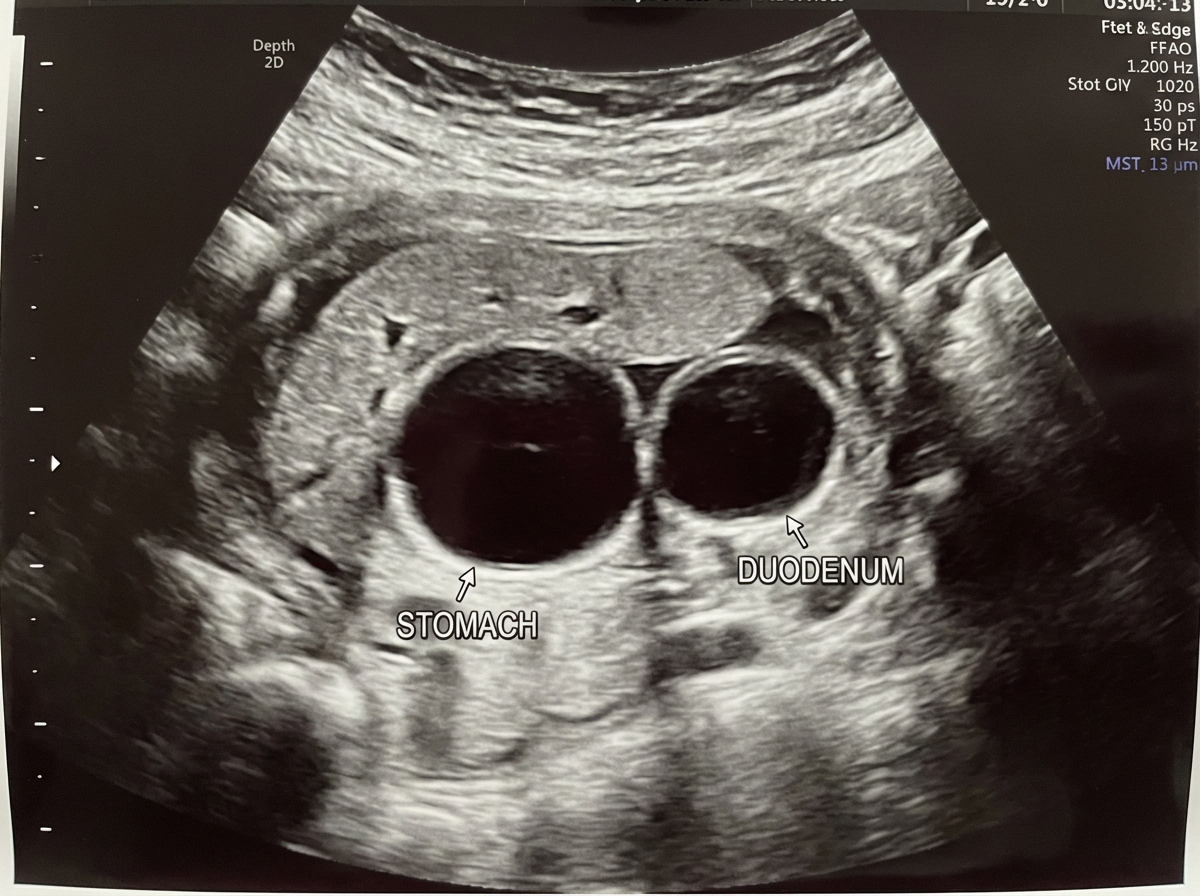
In which genetic condition is this most likely to be seen?
Which of the following is NOT a potential fetal injury in breech extraction?
In which of the following situations should forceps not be used for delivery?
Why is a transverse uterine incision preferred over a vertical incision during surgery?
All the following are criteria for outlet forceps application EXCEPT?
What procedure should be performed in case of arrest of the aftercoming head due to a contracted pelvis in a breech presentation?
External cephalic version is contraindicated in all of the following conditions except:
What is a chignon?
Which is the commonest indication for a classical cesarean section?
Practice by Chapter
Cesarean Section Techniques
Practice Questions
Vaginal Birth After Cesarean
Practice Questions
Instrumental Deliveries
Practice Questions
Breech Delivery
Practice Questions
Episiotomy and Repair
Practice Questions
Management of Multiple Gestation
Practice Questions
Cervical Cerclage
Practice Questions
Obstetric Hysterectomy
Practice Questions
Surgery During Pregnancy
Practice Questions
Surgical Complications in Obstetrics
Practice Questions
Want unlimited practice?
Get full access to all questions, explanations, and performance tracking.
Scan to download app