Vaginal defense is lost:
Which of the following is NOT a common complication in a neonate born to a mother with diabetes mellitus?
What are the characteristics after 28 weeks of gestation?
Low birth weight baby is defined as a baby weighing:
A baby is born with a large defect in the occipital bone through which the posterior portion of the brain has herniated. Which of the following terms best describes this lesion?
During a clinical examination, a senior resident asks an intern to examine the umbilical cord. How many arteries and veins are normally present in a healthy umbilical cord?
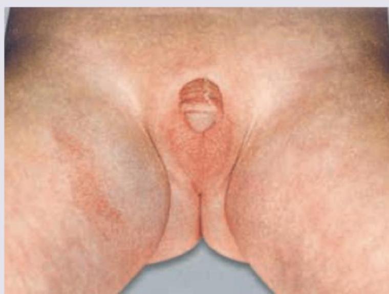
A Lethargic hypoglycemic girl neonate has the following genital appearance. What is the probable cause? (Recent Neet Pattern 2016-17)

Which of the following can be a complication in the baby due to post maturity of pregnancy ?
Meconium aspiration syndrome can be prevented by taking the following measures except :
A woman suffering from active tuberculosis not on ATT has a full term vaginal delivery. All the following should be done except:
Explanation: **Explanation:** The vaginal defense mechanism is primarily dependent on the **vaginal pH** and the presence of **Doderlein’s bacilli** (Lactobacilli). This environment is regulated by the levels of circulating maternal estrogen. 1. **Why Option B is Correct:** At birth, a female neonate has high levels of maternal estrogen in her circulation. This causes the vaginal epithelium to be thick, rich in glycogen, and acidic (pH 4–5) due to the presence of Doderlein’s bacilli. However, maternal estrogen is cleared from the infant's system within a few days. By **after 10 days of birth**, the estrogen levels drop significantly, the glycogen disappears, the pH becomes neutral or alkaline (pH 7), and Doderlein’s bacilli vanish. This loss of acidity results in the loss of the natural vaginal defense, making the infant susceptible to infection (vulvovaginitis) until puberty. 2. **Why Other Options are Incorrect:** * **Within 10 days of birth:** During the first 10 days, the "passive immunity" provided by maternal estrogen is still active; the vagina remains acidic and protected. * **During pregnancy:** Vaginal defense is actually **enhanced** during pregnancy due to high estrogen levels, which increase glycogen deposition and promote a highly acidic environment (pH 3.5–4.5) to protect against ascending infections. * **At puberty:** This is when vaginal defense is **re-established**. The surge in endogenous estrogen leads to the reappearance of glycogen and Doderlein’s bacilli, restoring the acidic pH. **High-Yield Clinical Pearls for NEET-PG:** * **Neonatal Period:** Vaginal pH is acidic (4–5) for the first 10 days, then becomes neutral (7) until puberty. * **Reproductive Age:** Vaginal pH is acidic (4–5.5). * **Menopause:** Vaginal pH becomes neutral or alkaline (6–7) due to estrogen deficiency. * **Doderlein’s Bacilli:** These convert glycogen into lactic acid, maintaining the protective acidic environment.
Explanation: In infants of diabetic mothers (IDM), the primary pathophysiology is **maternal hyperglycemia** leading to **fetal hyperinsulinemia**. **Why Hypercalcemia is the Correct Answer:** Neonates born to diabetic mothers typically experience **Hypocalcemia**, not hypercalcemia. This occurs because maternal diabetes is often associated with maternal magnesium loss, leading to fetal hypomagnesemia. Low magnesium levels suppress the secretion of Parathyroid Hormone (PTH) in the neonate, resulting in transient hypocalcemia within the first 24–72 hours of life. **Analysis of Incorrect Options:** * **Hypoglycemia:** This is the most common metabolic complication. High maternal glucose crosses the placenta, stimulating fetal pancreatic beta cells. After birth, the glucose supply is cut off, but the neonate’s hyperinsulinemia persists, causing a rapid drop in blood sugar. * **Hypokalemia:** This occurs secondary to hyperinsulinemia. Insulin shifts potassium from the extracellular space into the cells. * **Obesity:** IDMs are often macrosomic (Large for Gestational Age) due to the anabolic effects of insulin. These children have a significantly higher risk of developing childhood obesity and Type 2 Diabetes later in life. **High-Yield Clinical Pearls for NEET-PG:** * **Most common cardiac anomaly:** Ventricular Septal Defect (VSD). * **Most specific cardiac anomaly:** Transposition of the Great Arteries (TGA). * **Most characteristic anomaly:** Caudal Regression Syndrome (Sacral Agenesis). * **Other complications:** Polycythemia (due to fetal hypoxia), Hyperbilirubinemia, and Respiratory Distress Syndrome (insulin inhibits surfactant production by antagonizing cortisol).
Explanation: This question addresses the physiological and legal milestones of fetal development at the transition into the third trimester. **Explanation of the Correct Answer:** By **28 weeks of gestation**, several critical developmental milestones are reached, making **Option D** the correct choice: 1. **Viability (Option A):** In many clinical guidelines and legal frameworks (including historical WHO definitions), 28 weeks is considered the threshold of "legal viability." While modern NICU care allows survival at earlier gestations, 28 weeks marks a significant increase in the probability of survival outside the womb. 2. **Weight (Option B):** According to standard intrauterine growth curves, the average fetal weight at 28 weeks is approximately **1000 to 1100 grams**. In the context of the Indian Neonatal and Obstetric guidelines, a birth weight of ≥1000g often defines a "viable" birth. 3. **Type II Pneumocytes (Option C):** While Type II pneumocytes begin to appear around 20–24 weeks, they become functionally significant and start producing **surfactant** in adequate amounts by 28 weeks. This maturation of the alveolar lining is what allows for gas exchange and prevents alveolar collapse, facilitating extrauterine life. **Why other options are considered together:** Since all three criteria—legal/clinical viability, a weight threshold of 1000g, and the functional presence of surfactant-producing cells—are characteristic of the 28-week milestone, "All of the above" is the most accurate description. **High-Yield Clinical Pearls for NEET-PG:** * **Definition of Stillbirth (India):** Birth of a fetus after 28 weeks (or >1000g) that shows no signs of life. * **L/S Ratio:** At 28 weeks, the Lecithin/Sphingomyelin ratio begins to rise, but typically reaches the "mature" level of 2:1 at **34–35 weeks**. * **Surfactant:** Composed primarily of Dipalmitoylphosphatidylcholine (DPPC). Its production is accelerated by maternal corticosteroid administration (Betamethasone/Dexamethasone).
Explanation: ### Explanation The classification of birth weight is a fundamental concept in perinatology, used globally to predict neonatal morbidity and mortality. **1. Why the Correct Answer is Right:** According to the **World Health Organization (WHO)**, a **Low Birth Weight (LBW)** baby is defined as a neonate whose birth weight is **less than 2500 grams (up to and including 2499 g)**, regardless of the gestational age. This cutoff is significant because infants weighing less than 2500g have a substantially higher risk of complications such as hypoglycemia, hypothermia, and respiratory distress. **2. Analysis of Incorrect Options:** * **A. 1000 gm:** This defines **Extremely Low Birth Weight (ELBW)**. These infants are at the highest risk and often require intensive NICU care. * **B. 1500 gm:** This defines **Very Low Birth Weight (VLBW)**. Babies in this category (1001g to 1500g) have significantly higher mortality rates than standard LBW babies. * **C. 2000 gm:** While clinically concerning, this does not represent a formal WHO classification threshold. **3. High-Yield Clinical Pearls for NEET-PG:** * **LBW Sub-classifications:** * **LBW:** < 2500 g * **VLBW:** < 1500 g * **ELBW:** < 1000 g * **Macrosomia:** Defined as a birth weight **> 4000 g** (some guidelines use > 4500 g), commonly associated with maternal diabetes. * **Small for Gestational Age (SGA):** Refers to a birth weight below the **10th percentile** for that specific gestational age. * **Ponderal Index:** Used to differentiate between symmetrical and asymmetrical IUGR. * **Incidence:** India has one of the highest incidences of LBW globally, primarily due to maternal malnutrition and anemia.
Explanation: ### Explanation **Correct Option: A. Encephalocele** An **encephalocele** is a neural tube defect (NTD) characterized by a sac-like protrusion of the brain and the membranes covering it (meninges) through an opening in the skull. In this case, the herniation of the "posterior portion of the brain" through an occipital bone defect fits the definition perfectly. The most common site for encephalocele is the **occipital region** (75% of cases). **Analysis of Incorrect Options:** * **B. Meningocele:** This refers to the herniation of the **meninges only** (dura and arachnoid) through a defect in the skull or vertebral column, without any neural tissue (brain or spinal cord) inside the sac. * **C. Myelocele:** This is a type of *Spina Bifida Cystica* where the spinal cord is exposed to the exterior, but it specifically involves the **spinal column**, not the cranium/occipital bone. * **D. Spina bifida:** This is a broad term for NTDs involving the **vertebral column** (spinal arches). It does not describe defects in the occipital bone or herniation of brain tissue. **NEET-PG High-Yield Pearls:** 1. **Etiology:** Like most NTDs, encephaloceles are associated with **folic acid deficiency** during the first trimester. 2. **Diagnosis:** Elevated **Alpha-fetoprotein (AFP)** in maternal serum and amniotic fluid is a key screening marker. 3. **Meckel-Gruber Syndrome:** A high-yield triad to remember is **Occipital Encephalocele + Polydactyly + Polycystic Kidneys**. 4. **Prognosis:** Occipital encephaloceles often contain the visual cortex or cerebellum; the prognosis depends on the amount of brain tissue herniated and the presence of associated hydrocephalus.
Explanation: ***1 vein and 2 arteries*** - The normal configuration of a healthy umbilical cord is three vessels: **two umbilical arteries** and **one umbilical vein**. - The **umbilical vein** transports oxygenated blood from the placenta to the fetus, whereas the **two umbilical arteries** carry deoxygenated blood and metabolic waste from the fetus back to the placenta. *1 artery and 2 veins* - This configuration is incorrect; normally, the right umbilical vein **atrophies** early in gestation, leaving only a single left umbilical vein. - A cord with two veins suggests a **developmental anomaly** and is not the standard finding. *1 artery and 1 vein* - This pattern is an anomaly known as a **Single Umbilical Artery (SUA)**, which is found in 0.5% to 1% of pregnancies. - SUA is clinically significant as it is associated with an increased risk of **congenital anomalies**, especially renal, cardiac, and chromosomal defects. *2 arteries and 2 veins* - This is an abnormal configuration; while two arteries are standard, the presence of two veins instead of one is due to the failure of the **right umbilical vein** to involute, which is typically a normal event. - The persistence of two veins is highly **atypical** and often requires specialized fetal surveillance.
Explanation: ***Congenital adrenal hyperplasia*** - The image shows **ambiguous genitalia (clitoromegaly/phallic structure with fused labia)** in a neonate presenting with **lethargy and hypoglycemia**, which are symptoms of **salt-wasting crisis**. - This clinical picture in a presumed genetic female is highly suggestive of **21-hydroxylase deficiency**, the most common form of CAH, leading to excess adrenal androgens and mineralocorticoid deficiency. *Ovotesticular disorder of sexual differentiation* - This condition involves the presence of both **ovarian and testicular tissue**, leading to ambiguous genitalia. - While it can cause ambiguous genitalia, it does not typically present with the acute metabolic crisis (hypoglycemia, lethargy) seen in this neonate, which points to a hormonal deficiency. *Turner syndrome* - **Turner syndrome (45, XO)** affects genetic females and is characterized by the absence of one X chromosome, typically resulting in **female external genitalia** that are usually normal or immature. - It does not cause ambiguous genitalia or the metabolic derangements (hypoglycemia) characteristic of adrenal insufficiency. *Klinefelter syndrome* - **Klinefelter syndrome (47, XXY)** affects genetic males and typically presents with **normal male external genitalia** at birth. - Features like hypogonadism and gynecomastia become apparent later in puberty, and it is not associated with ambiguous genitalia or neonatal metabolic crises.
Explanation: ***Meconium aspiration*** - **Post-term pregnancies** (>42 weeks) are associated with **oligohydramnios** and **placental insufficiency**, leading to fetal distress - Fetal distress causes passage of **meconium** into amniotic fluid, which can be aspirated during gasping movements - **Meconium aspiration syndrome** is one of the **most characteristic complications** of post-maturity - This is the **most recognized** complication among the options listed *Hypoglycemia* - Post-term infants **ARE actually at risk** for hypoglycemia - Mechanism: **Placental insufficiency** leads to depleted fetal **glycogen stores** and subcutaneous fat - However, this is **less specific** to post-term pregnancy compared to meconium aspiration, as it occurs in multiple conditions - While medically correct, meconium aspiration is the more characteristic complication *Intraventricular hemorrhage* - This is primarily a complication of **prematurity**, especially in very low birth weight infants - Caused by fragility of the **germinal matrix** in preterm brains - **Not associated with post-term pregnancy** *Polycythemia* - Post-term infants **can develop polycythemia** (hematocrit >65%) - Mechanism: Chronic **placental insufficiency** → fetal hypoxia → increased **erythropoietin production** - While this is a recognized complication, **meconium aspiration** remains the **most classic and frequently tested** complication of post-maturity
Explanation: ***Delivering the baby by emergency caesarean section*** - While **emergency caesarean section** may be indicated for fetal distress in the setting of meconium-stained amniotic fluid, it does **not directly prevent meconium aspiration syndrome**. - If the fetus has already passed meconium and gasping movements have occurred in utero, aspiration may have already happened before delivery regardless of the mode of delivery. - Emergency delivery addresses the underlying fetal compromise but is not a specific preventive measure for meconium aspiration. *Amnioinfusion* - **Amnioinfusion** is a proven preventive measure that involves transcervical infusion of normal saline into the uterine cavity to dilute thick meconium-stained amniotic fluid. - This reduces meconium concentration and viscosity, decreasing the risk and severity of meconium aspiration syndrome. - Multiple studies have demonstrated that amnioinfusion can reduce the incidence of severe MAS and improve neonatal outcomes. *Oropharyngeal suctioning* - **Note:** Current neonatal resuscitation guidelines (2015 onwards) have de-emphasized routine intrapartum oropharyngeal and nasopharyngeal suctioning. - Historically, this was thought to prevent aspiration, but recent evidence shows **routine suctioning does not reduce the incidence of MAS** and may delay initiation of ventilation. - However, in the context of this historical exam question (UPSC-CMS-2013), this was considered a preventive measure. *Suctioning of trachea through laryngoscope* - **Note:** Current guidelines (2015 onwards) **no longer recommend routine endotracheal suctioning** for non-vigorous infants born through meconium-stained amniotic fluid. - Studies showed that routine tracheal suctioning did not improve outcomes and delayed initiation of positive pressure ventilation. - Current practice prioritizes **immediate initiation of ventilation** for non-vigorous newborns rather than routine suctioning. - In the historical context of this 2013 exam, tracheal suctioning was still considered standard practice for prevention.
Explanation: ***BCG should be given to the neonate*** - This is the **EXCEPTION** - BCG vaccination should be **deferred** in neonates born to mothers with active, untreated tuberculosis. - **Current guidelines (WHO/CDC):** BCG is contraindicated or delayed when there is known exposure to active TB, as the neonate is at risk of developing active disease, and BCG given during the incubation period may cause disseminated BCG infection. - The neonate should first receive **INH prophylaxis for 6 months**, then be reassessed. If TB is ruled out and tuberculin skin test is negative, BCG can be given after completing prophylaxis. - Immediate BCG vaccination in an exposed neonate may lead to complications if the infant develops active TB. *Breastfeed the neonate* - **Breastfeeding should be continued** even when the mother has active tuberculosis, as per WHO and CDC recommendations. - **TB is NOT transmitted through breast milk** - the infection spreads via respiratory droplets, not through lactation. - The mother should wear a **surgical mask during breastfeeding** and feeding times to minimize respiratory transmission risk. - The benefits of breastfeeding (nutrition, immunity, bonding) outweigh the risks when appropriate precautions are taken. *Neonate should be isolated from mother* - **Temporary separation is recommended** until the mother with active, untreated TB is rendered non-infectious. - Isolation prevents **respiratory transmission** of Mycobacterium tuberculosis from mother to the highly susceptible neonate. - Once the mother has been on effective anti-tuberculosis treatment (ATT) for **at least 2 weeks** and shows clinical improvement, contact can be resumed with appropriate precautions (mask use). *Neonate should be given INH* - **Isoniazid (INH) prophylaxis is mandatory** for neonates born to mothers with active tuberculosis. - Standard protocol: **INH 10 mg/kg/day for 6 months** to prevent progression from latent to active TB. - After 6 months of prophylaxis, the infant should be evaluated with tuberculin skin test (TST). If negative and no evidence of active disease, INH can be stopped and BCG can be administered. - This prophylactic approach significantly reduces the risk of the neonate developing active tuberculosis.
Neonatal Resuscitation
Practice Questions
Care of the Normal Newborn
Practice Questions
Low Birth Weight and Prematurity
Practice Questions
Neonatal Infections
Practice Questions
Birth Asphyxia and Hypoxic-Ischemic Encephalopathy
Practice Questions
Neonatal Jaundice
Practice Questions
Respiratory Distress in Newborn
Practice Questions
Congenital Anomalies
Practice Questions
Birth Injuries
Practice Questions
Perinatal Mortality and Morbidity
Practice Questions
Get full access to all questions, explanations, and performance tracking.
Start For Free