
Neonatology and Perinatology — MCQs

On this page
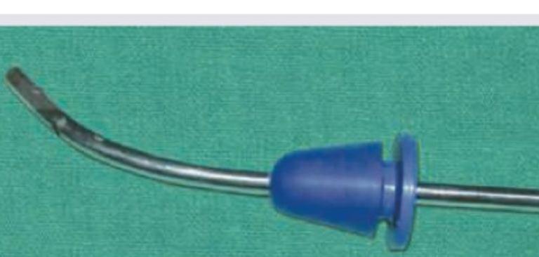
Q1
Identify the instrument:
Practice by Chapter
1
Neonatal Resuscitation
Practice Questions
2
Care of the Normal Newborn
Practice Questions
3
Low Birth Weight and Prematurity
Practice Questions
4
Neonatal Infections
Practice Questions
5
Birth Asphyxia and Hypoxic-Ischemic Encephalopathy
Practice Questions
6
Neonatal Jaundice
Practice Questions
7
Respiratory Distress in Newborn
Practice Questions
8
Congenital Anomalies
Practice Questions
9
Birth Injuries
Practice Questions
10
Perinatal Mortality and Morbidity
Practice Questions
Want unlimited practice?
Get full access to all questions, explanations, and performance tracking.
Scan to download app