
Maternal-Fetal Medicine — MCQs

On this page
A 30-year-old G1P1001 patient presents at 37 weeks gestational age with a breech presentation. The cervix is 50% effaced and 1-2 cm dilated. The estimated fetal weight is 7 lbs. The patient is offered all the following management plans EXCEPT:
A pregnant woman at 39 weeks gestation with a singleton fetus in cephalic presentation develops chickenpox. She has no other pregnancy complications. What is the best method to prevent neonatal infection?
Singer's alkali denaturation test is performed for?
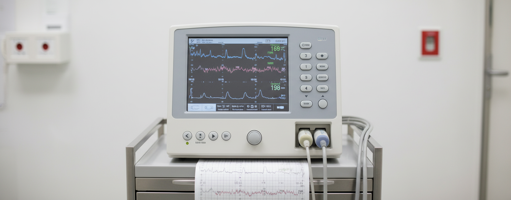
The above shown device is used for:
After 20 weeks of gestation, what is the main contribution to amniotic fluid?
Hydramnios is complicated by which of the following conditions, EXCEPT?
Abruptio placentae occurs in all except?
Magnesium sulphate toxicity includes all of the following EXCEPT?
All of the following are true about SLE in pregnancy except?
Which of the following is NOT a risk factor for preterm delivery?
Practice by Chapter
Fetal Assessment Techniques
Practice Questions
Hypertensive Disorders in Pregnancy
Practice Questions
Intrauterine Growth Restriction
Practice Questions
Multiple Gestation
Practice Questions
Rh Isoimmunization and Other Blood Group Incompatibilities
Practice Questions
Intrauterine Fetal Therapy
Practice Questions
Prenatal Diagnosis and Genetic Counseling
Practice Questions
Placental Abnormalities
Practice Questions
Preterm Labor and Delivery
Practice Questions
Management of Medical Disorders in Pregnancy
Practice Questions
Want unlimited practice?
Get full access to all questions, explanations, and performance tracking.
Scan to download app