
Maternal-Fetal Medicine — MCQs

On this page
Early diastolic notching of the uterine artery disappears by which week of gestation?
A 22-year-old woman presents with sudden onset of severe lower abdominal pain. Physical examination reveals no masses but severe tenderness in the right lower quadrant. Pelvic examination shows no lesions of the cervix or vagina. Bowel sounds are present. An abdominal ultrasound scan shows a 4-cm focal enlargement of the proximal right fallopian tube. A dilation and curettage procedure reveals decidua only from the endometrial cavity. Which of the following laboratory findings is most likely to be reported for this patient?
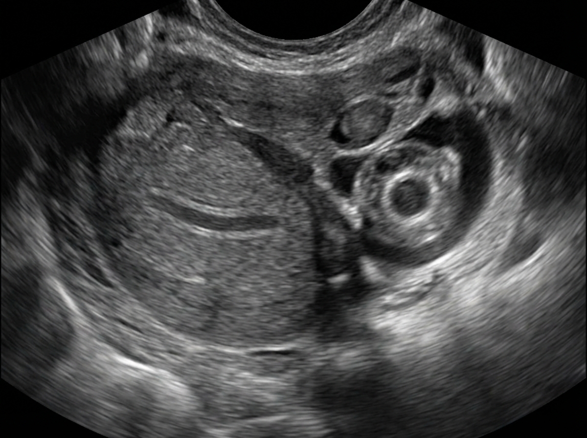
What does the following ultrasound show?
What is the drug of choice for the prevention of seizures in a patient with severe preeclampsia?
In post-term pregnancy, what is the increased risk for the fetus, excluding one option?
What is the treatment of choice for intrahepatic cholestasis of pregnancy?
What is the earliest fetal anomaly detectable by ultrasound?
A 32-year-old primi gravida presents with deep vein thrombosis (DVT). There is a history of two episodes of DVT in the past and she was diagnosed to have antiphospholipid antibody syndrome. What is the next step in management?
A 35-year-old lady, G4 P3, presents with amenorrhea of 12 weeks, excessive vomiting, and bleeding per vaginam. On examination, her pulse is 90/min, BP is 150/110 mm Hg, and the uterus is of 20 weeks size on palpation. What is your most probable diagnosis?
A 28-week pregnant patient presents with abdominal pain and fever. Ultrasonography reveals the presence of a fibroid. What is the next line of management?
Practice by Chapter
Fetal Assessment Techniques
Practice Questions
Hypertensive Disorders in Pregnancy
Practice Questions
Intrauterine Growth Restriction
Practice Questions
Multiple Gestation
Practice Questions
Rh Isoimmunization and Other Blood Group Incompatibilities
Practice Questions
Intrauterine Fetal Therapy
Practice Questions
Prenatal Diagnosis and Genetic Counseling
Practice Questions
Placental Abnormalities
Practice Questions
Preterm Labor and Delivery
Practice Questions
Management of Medical Disorders in Pregnancy
Practice Questions
Want unlimited practice?
Get full access to all questions, explanations, and performance tracking.
Scan to download app