Oligohydramnios is associated with which of the following conditions?
In a patient with Antiphospholipid Antibody syndrome, which of the following treatments is used to prevent further abortions?
What is the investigation of choice for an ectopic pregnancy?
Large placenta is seen in all of the following conditions except?
Doppler ultrasonography in Intrauterine Growth Restriction (IUGR) and Preeclampsia shows a notch in which artery?
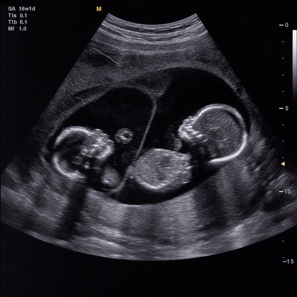
A 26-year-old primigravida with a twin gestation at 30 weeks presents for a USG. The sonogram indicates that the fetuses are both male and the placenta appears to be diamniotic and monochorionic. Twin B is noted to have oligohydramnios and to be much smaller than twin A. In this clinical scenario, all of the following are concerns for twin A except?
Velamentous insertion of the umbilical cord is associated with an increased risk for which of the following?
What is the appropriate management of vulvar varices during pregnancy?
What is the most common cause of fulminant hepatitis in pregnancy?
A pregnant woman, G2P1, at 35 weeks gestation complains of decreased fetal movement. What is the next step in management?
Explanation: **Explanation:** **Oligohydramnios** is defined as an Amniotic Fluid Index (AFI) < 5 cm or a Single Deepest Pocket (SDP) < 2 cm. The volume of amniotic fluid is primarily maintained by a balance between fetal urine production (major source) and fetal swallowing (major clearance). **Why Renal Agenesis is Correct:** In the second and third trimesters, fetal urine is the primary contributor to amniotic fluid volume. In **Renal Agenesis** (Potter’s Sequence), the absence of kidneys leads to a failure of urine production (**anuria**). This results in severe oligohydramnios, which subsequently causes pulmonary hypoplasia, limb deformities, and characteristic facial features due to uterine pressure. **Analysis of Incorrect Options:** * **B. Neural Tube Defects (NTDs):** These are typically associated with **Polyhydramnios**. In open NTDs (like anencephaly), there is excessive transudation of fluid from the exposed meninges and a lack of fetal swallowing coordination. * **C. Postmature Birth:** While post-term pregnancy (>42 weeks) is a known cause of oligohydramnios due to placental insufficiency and reduced fetal renal perfusion, "Postmature birth" is a general term. In the context of this question, **Renal Agenesis** is the classic, definitive pathological association. * **D. Premature Birth:** Prematurity itself does not cause oligohydramnios. However, Preterm Premature Rupture of Membranes (PPROM) can lead to low fluid, but the birth is a result, not the primary cause of the fluid status. **High-Yield Clinical Pearls for NEET-PG:** * **Most common cause of Oligohydramnios:** Premature Rupture of Membranes (PROM). * **Most common congenital anomaly causing it:** Renal anomalies (Posterior Urethral Valves in males, Bilateral Renal Agenesis). * **Drug-induced Oligohydramnios:** ACE inhibitors (cause fetal renal tubular dysgenesis) and NSAIDs (reduce fetal urine output). * **Amniotic Band Syndrome:** A serious complication of early, severe oligohydramnios.
Explanation: **Explanation:** Antiphospholipid Antibody Syndrome (APS) is an autoimmune condition characterized by recurrent pregnancy loss and/or thromboembolism. In pregnancy, APS causes placental insufficiency through thrombosis of the uteroplacental vasculature and direct inhibition of trophoblastic invasion. **1. Why Option A is Correct:** The standard of care for preventing recurrent abortions in APS is the combination of **Low-Dose Aspirin (LDA)** and **Low Molecular Weight Heparin (LMWH)**. * **Aspirin (75–150 mg):** Inhibits thromboxane A2, reducing platelet aggregation and improving placental perfusion. * **LMWH (Prophylactic dose):** Prevents thrombosis and has anti-inflammatory properties that aid trophoblast implantation. Combined therapy is significantly more effective than aspirin alone, achieving a live birth rate of approximately 70–80%. **2. Why Other Options are Incorrect:** * **Corticosteroids (B):** Previously used to suppress antibodies, but they are no longer recommended as primary therapy because they increase the risk of gestational diabetes, hypertension, and preterm birth without improving live birth rates. * **IVIG (C) and Plasmapheresis (D):** These are considered "rescue therapies" reserved for **Refractory APS** (failure of Heparin + Aspirin) or **Catastrophic APS**. They are not first-line treatments due to high cost and lack of superior efficacy in routine cases. **3. High-Yield Clinical Pearls for NEET-PG:** * **Diagnosis:** Requires at least one clinical criteria (Vascular thrombosis or Pregnancy morbidity) AND one laboratory criteria (Lupus anticoagulant, Anti-cardiolipin, or Anti-β2 glycoprotein-I) positive on two occasions, 12 weeks apart. * **Pregnancy Morbidity Definition:** ≥3 unexplained consecutive abortions (<10 weeks), ≥1 death of a morphologically normal fetus (≥10 weeks), or ≥1 preterm birth (<34 weeks) due to severe preeclampsia/placental insufficiency. * **Warfarin Warning:** Warfarin is contraindicated in pregnancy (teratogenic); patients on long-term warfarin must switch to LMWH before conception or by 6 weeks gestation.
Explanation: **Explanation:** The investigation of choice for diagnosing an ectopic pregnancy is **Transvaginal Ultrasonography (TVUS)**. **Why Transvaginal USG is the Correct Answer:** TVUS is the gold standard because it offers superior resolution compared to transabdominal scans, allowing for the visualization of an intrauterine gestational sac as early as 4.5 to 5 weeks. The definitive diagnosis of an ectopic pregnancy is made when an extrauterine gestational sac with a yolk sac or embryo (with or without cardiac activity) is visualized. The most common finding, however, is an adnexal mass separate from the ovary (the "tubal ring" sign). **Analysis of Incorrect Options:** * **CT Scan:** Not used in pregnancy due to ionizing radiation and poor soft-tissue resolution for early pelvic structures. * **Serum hCG levels:** While crucial for management, a single hCG level cannot diagnose the *location* of a pregnancy. It is used in conjunction with TVUS to determine the "Discriminatory Zone" (usually 1500–2000 mIU/mL), above which an intrauterine sac should be visible. * **MRI:** Highly accurate but expensive and time-consuming; it is reserved for rare, complex cases like abdominal or interstitial pregnancies when USG is inconclusive. **High-Yield Clinical Pearls for NEET-PG:** * **Most common site:** Ampulla of the Fallopian tube. * **Most common USG finding:** An adnexal mass (non-specific) or the "Bagel Sign" (tubal ring). * **Pseudosac:** A midline fluid collection in the uterus seen in 10% of ectopic cases; unlike a true gestational sac, it lacks a double decidual sign. * **Gold Standard for Diagnosis:** Laparoscopy (though TVUS is the investigation of choice).
Explanation: **Explanation:** The size and weight of the placenta are generally proportional to the size of the fetus. A **Large Placenta (Placentomegaly)** is typically defined as a placental thickness >4 cm or a weight exceeding the 90th percentile for gestational age. **1. Why IUGR is the correct answer:** In **Intrauterine Growth Restriction (IUGR)**, particularly placental-mediated IUGR (Type 2/Asymmetrical), there is **uteroplacental insufficiency**. This is characterized by poor trophoblastic invasion and reduced surface area for nutrient exchange, leading to a **small, shrunken, and infarcted placenta**. Since the question asks for the condition where a large placenta is *not* seen, IUGR is the correct choice. **2. Why the other options are incorrect:** * **Syphilis:** Congenital syphilis causes chronic villitis and edema of the placental villi, leading to a significantly enlarged, "pale," and "greasy" placenta. * **Cytomegalovirus (CMV) & Rubella:** These are part of the **TORCH infections**. Chronic intrauterine infections lead to inflammatory infiltration (villitis) and compensatory hyperplasia of the placental tissue, resulting in placentomegaly. **NEET-PG High-Yield Pearls:** * **Causes of Large Placenta:** Diabetes Mellitus (most common), Hydrops Fetalis (Immune/Non-immune), Syphilis, TORCH infections, Multiple gestations, and Chorioangioma. * **Causes of Small Placenta:** IUGR, Pre-eclampsia, Chronic Hypertension, and Chromosomal anomalies (e.g., Trisomy 18). * **Normal Placental Weight:** Approximately 1/6th of the baby's weight at term (approx. 500g). * **Placentomegaly on Ultrasound:** A thickness of **>40 mm** is a key diagnostic marker.
Explanation: **Explanation:** The correct answer is **B. Uterine artery**. In a normal pregnancy, trophoblastic invasion of the spiral arteries converts them from high-resistance, low-flow vessels into low-resistance, high-flow vessels. This process occurs in two waves (at 10–12 weeks and 16–20 weeks). If this invasion is defective, as seen in **Preeclampsia and Fetal Growth Restriction (FGR/IUGR)**, the uterine artery remains a high-resistance vessel. On Doppler, this high resistance manifests as an **early diastolic notch**. Persistence of this notch beyond **24 weeks** of gestation is a significant predictor of placental insufficiency, preeclampsia, and IUGR. **Analysis of Incorrect Options:** * **A. Umbilical artery:** Doppler changes here reflect fetal-placental resistance. In IUGR, we see increased resistance, reduced end-diastolic flow (REDF), or absent/reversed end-diastolic flow (AEDF/REDF), but **not** a diastolic notch. * **C. Internal iliac artery:** While the uterine artery is a branch of the internal iliac, the specific pathological changes related to placental perfusion are measured directly at the uterine artery. * **D. Vitelline artery:** This is an embryonic vessel associated with the yolk sac and is not used for monitoring IUGR or preeclampsia in clinical practice. **High-Yield Clinical Pearls for NEET-PG:** * **Uterine Artery Doppler:** Best for **screening** and predicting preeclampsia (High resistance = Notch). * **Umbilical Artery Doppler:** Best for **monitoring** a diagnosed case of IUGR and deciding the timing of delivery. * **Middle Cerebral Artery (MCA):** Shows "Brain Sparing Effect" (increased diastolic flow/low resistance) in IUGR. * **Ductus Venosus:** Abnormal flow (a-wave reversal) is a late sign of fetal cardiac failure and an indication for immediate delivery.
Explanation: This question describes a classic case of **Twin-to-Twin Transfusion Syndrome (TTTS)**, which occurs in monochorionic (MC) diamniotic twin gestations due to unbalanced vascular anastomoses (specifically deep arteriovenous anastomoses) in the shared placenta. ### **Why Anemia is the Correct Answer** In TTTS, blood is shunted from the "Donor" twin to the "Recipient" twin. * **Twin B (Donor):** Becomes anemic, growth-restricted, and develops oligohydramnios due to decreased renal perfusion. * **Twin A (Recipient):** Receives excess blood volume, leading to **polycythemia** (not anemia), hypervolemia, and polyuria (hydramnios). Therefore, anemia is a concern for Twin B, while polycythemia is the concern for Twin A. ### **Explanation of Incorrect Options** * **A. Congestive Heart Failure (CHF):** The recipient twin (Twin A) suffers from chronic volume overload. This leads to cardiac hypertrophy, cardiomegaly, and eventually high-output heart failure and hydrops fetalis. * **C. Hydramnios:** Excess blood volume in the recipient twin leads to increased glomerular filtration and fetal polyuria, resulting in polyhydramnios (the "Poly" in the Poly-Oli sequence). * **D. Widespread Thromboses:** If one twin dies in utero, the surviving twin (Twin A) is at high risk for thrombotic events, DIC, and multi-organ ischemia (e.g., multicystic encephalomalacia) due to the transfer of thromboplastic material or sudden hemodynamic shifts through placental anastomoses. ### **High-Yield Clinical Pearls for NEET-PG** * **Diagnosis:** TTTS is diagnosed via USG showing a single placenta, same-sex twins, and the **"Poly-Oli sequence"** (Deepest Vertical Pocket >8cm in one sac and <2cm in the other). * **Staging:** Uses the **Quintero Staging System** (Stage I: Poly/Oli; Stage II: Absent bladder in donor; Stage III: Abnormal Dopplers; Stage IV: Hydrops; Stage V: Death). * **Treatment of Choice:** Fetoscopic **Laser Ablation** of the placental anastomoses (most effective between 16–26 weeks). Serial amnioreduction is a palliative alternative.
Explanation: **Explanation:** **Velamentous insertion of the umbilical cord** occurs when the umbilical vessels separate in the membranes at a distance from the placental margin, traveling between the amnion and chorion without the protection of **Wharton’s jelly**. **Why Option B is Correct:** Because the vessels are unprotected, they are highly susceptible to compression or rupture. If these vessels overlie the internal os (**Vasa Previa**), they can rupture during spontaneous or artificial rupture of membranes. Since the blood within these vessels is of **fetal origin**, even a small amount of bleeding can lead to rapid **fetal exsanguination** and death, often occurring before or during early labor. **Analysis of Incorrect Options:** * **Option A:** The bleeding in velamentous insertion/vasa previa is fetal, not maternal. While maternal hemorrhage (like Abruptio Placentae) is a differential for antepartum hemorrhage, it is not the primary risk here. * **Option C:** While cord torsion can occur in any pregnancy, it is more commonly associated with a long umbilical cord or hypercoiled cord, not specifically velamentous insertion. * **Option D:** Velamentous insertion is associated with placenta previa, multiple gestations, and IUGR, but it is not a primary cause of structural fetal malformations. **High-Yield Clinical Pearls for NEET-PG:** * **Vasa Previa Triad:** Rupture of membranes + Painless vaginal bleeding + Fetal bradycardia/distress. * **Apt Test / Alkali Denaturation Test:** Used to differentiate fetal hemoglobin (HbF) from maternal hemoglobin (HbA) in vaginal blood. HbF remains pink (resistant to alkali), while HbA turns yellow-brown. * **Association:** Velamentous insertion is significantly more common in **monochorionic twin gestations** and pregnancies resulting from **IVF**.
Explanation: **Explanation:** **Vulvar varices** occur in approximately 4–10% of pregnancies, typically due to increased pelvic venous pressure, progesterone-induced vasodilation, and the compressive effect of the gravid uterus on the inferior vena cava. **Why "Observation only" is correct:** The management of vulvar varices during pregnancy is almost exclusively **conservative (observation)**. The key clinical concept is that these varicosities are physiological in nature and **spontaneously regress** within 6–8 weeks postpartum once the mechanical obstruction (the fetus) is removed and hormonal levels normalize. Active surgical or invasive intervention is avoided during pregnancy due to the high risk of bleeding and the likelihood of natural resolution. **Why other options are incorrect:** * **Pressure:** While support garments or "vulvar trusses" can provide symptomatic relief for pain or heaviness, they do not treat the underlying condition. * **Cautery:** This is contraindicated as it carries a high risk of severe hemorrhage from the engorged, thin-walled veins and does not address the proximal venous congestion. * **Simple vulvectomy:** This is a radical surgical procedure reserved for malignancies; it is never indicated for benign gestational varicosities. **NEET-PG High-Yield Pearls:** * **Mode of Delivery:** Vulvar varices are **not** an indication for Cesarean section. Vaginal delivery is safe as these veins are low-pressure and rarely rupture during birth. * **Episiotomy:** If a vaginal delivery is performed, avoid an episiotomy if possible to prevent accidental injury to a varix, which can lead to significant bleeding. * **Symptoms:** Most patients are asymptomatic, but some may report a "heavy" sensation or visible swelling that worsens with prolonged standing.
Explanation: ### Explanation **Correct Answer: D. Hepatitis E** **1. Why Hepatitis E is the Correct Answer:** Hepatitis E Virus (HEV), specifically genotypes 1 and 2, is the most common cause of fulminant hepatic failure (FHF) in pregnant women, particularly in developing countries like India. While HEV is typically a self-limiting infection in the general population (mortality <1%), it takes a devastating course during pregnancy, especially in the **third trimester**. The mortality rate can soar to **15–25%**. The underlying mechanism is believed to involve a combination of high viral load, altered maternal immune response (Th2 shift), and high levels of steroid hormones which enhance viral replication. **2. Why Other Options are Incorrect:** * **Hepatitis B (A):** While HBV is a common cause of chronic hepatitis and can cause acute liver failure, it is not the *most common* cause of fulminant hepatitis specifically triggered by the pregnant state. * **Hepatitis C (B):** HCV rarely causes acute fulminant hepatitis; it is primarily known for its progression to chronic liver disease and cirrhosis. * **Hepatitis D (C):** HDV only occurs as a co-infection or super-infection with HBV. While it increases the severity of HBV, it is not the leading cause of FHF in pregnancy. **3. Clinical Pearls for NEET-PG:** * **Transmission:** HEV is transmitted via the **fecal-oral route** (waterborne). * **Vertical Transmission:** HEV has a high rate of vertical transmission, often leading to neonatal hypoglycemia and jaundice. * **Differential Diagnosis:** Fulminant HEV must be distinguished from pregnancy-specific liver diseases like **Acute Fatty Liver of Pregnancy (AFLP)** and **HELLP syndrome**. * **Prognosis:** The most common cause of death in HEV-infected pregnant women is disseminated intravascular coagulation (DIC) and encephalopathy.
Explanation: ### Explanation **1. Why Non-stress Test (NST) is the Correct Answer:** Decreased fetal movement (DFM) is a clinical warning sign of potential fetal compromise. The primary goal in management is to assess fetal well-being promptly using the least invasive method. The **Non-stress Test (NST)** is the standard **first-line screening tool** for evaluating fetal heart rate (FHR) reactivity in the third trimester (after 28–32 weeks). A reactive NST (two or more accelerations in 20 minutes) indicates a functional fetal brainstem and an intact autonomic nervous system, effectively ruling out immediate acidemia. **2. Analysis of Incorrect Options:** * **A. Observation:** This is inappropriate and dangerous. DFM can be a precursor to stillbirth; therefore, objective fetal surveillance is mandatory. * **C. Contraction Stress Test (CST):** This is a second-line test used when the NST is non-reactive. It involves inducing contractions (via oxytocin or nipple stimulation) to check for late decelerations. It is more time-consuming and contraindicated in certain conditions (e.g., placenta previa). * **D. Biophysical Profile Score (BPS):** While BPS is a highly accurate predictor of fetal health, it is typically reserved as a **follow-up test** if the NST is non-reactive or if the patient is high-risk. It requires ultrasound equipment and more time than a bedside NST. **3. Clinical Pearls for NEET-PG:** * **Definition of DFM:** Usually defined as <10 movements in 2 hours (Sadovsky’s method) or <3 movements in 1 hour (Cardiff "Count-to-Ten"). * **Sequence of Assessment:** DFM → Clinical History/Auscultation → **NST** → (if non-reactive) → **BPS** or CST. * **Modified BPS:** Consists of NST + Amniotic Fluid Index (AFI). This is a common high-yield alternative for rapid assessment. * **Reactive NST:** 2 accelerations of 15 bpm lasting 15 seconds in a 20-minute window (15x15 rule). For <32 weeks, use the 10x10 rule.
Fetal Assessment Techniques
Practice Questions
Hypertensive Disorders in Pregnancy
Practice Questions
Intrauterine Growth Restriction
Practice Questions
Multiple Gestation
Practice Questions
Rh Isoimmunization and Other Blood Group Incompatibilities
Practice Questions
Intrauterine Fetal Therapy
Practice Questions
Prenatal Diagnosis and Genetic Counseling
Practice Questions
Placental Abnormalities
Practice Questions
Preterm Labor and Delivery
Practice Questions
Management of Medical Disorders in Pregnancy
Practice Questions
Get full access to all questions, explanations, and performance tracking.
Start For Free