
Maternal-Fetal Medicine — MCQs

On this page
A couple with an Rh-positive female and an Rh-negative male seeks counseling regarding pregnancy. What is the recommended course of action?
Which is the earliest ectopic pregnancy location to undergo rupture?
A 35 weeks pregnant woman has ultrasound parameters corresponding to gestational age. Doppler shows absent end diastolic flow. What is the recommended management?
Among the following, which is the most sensitive ultrasonographic finding of Trisomy 21?
Which of the following is NOT an explanation for decreased variability of the fetal heart rate tracing?
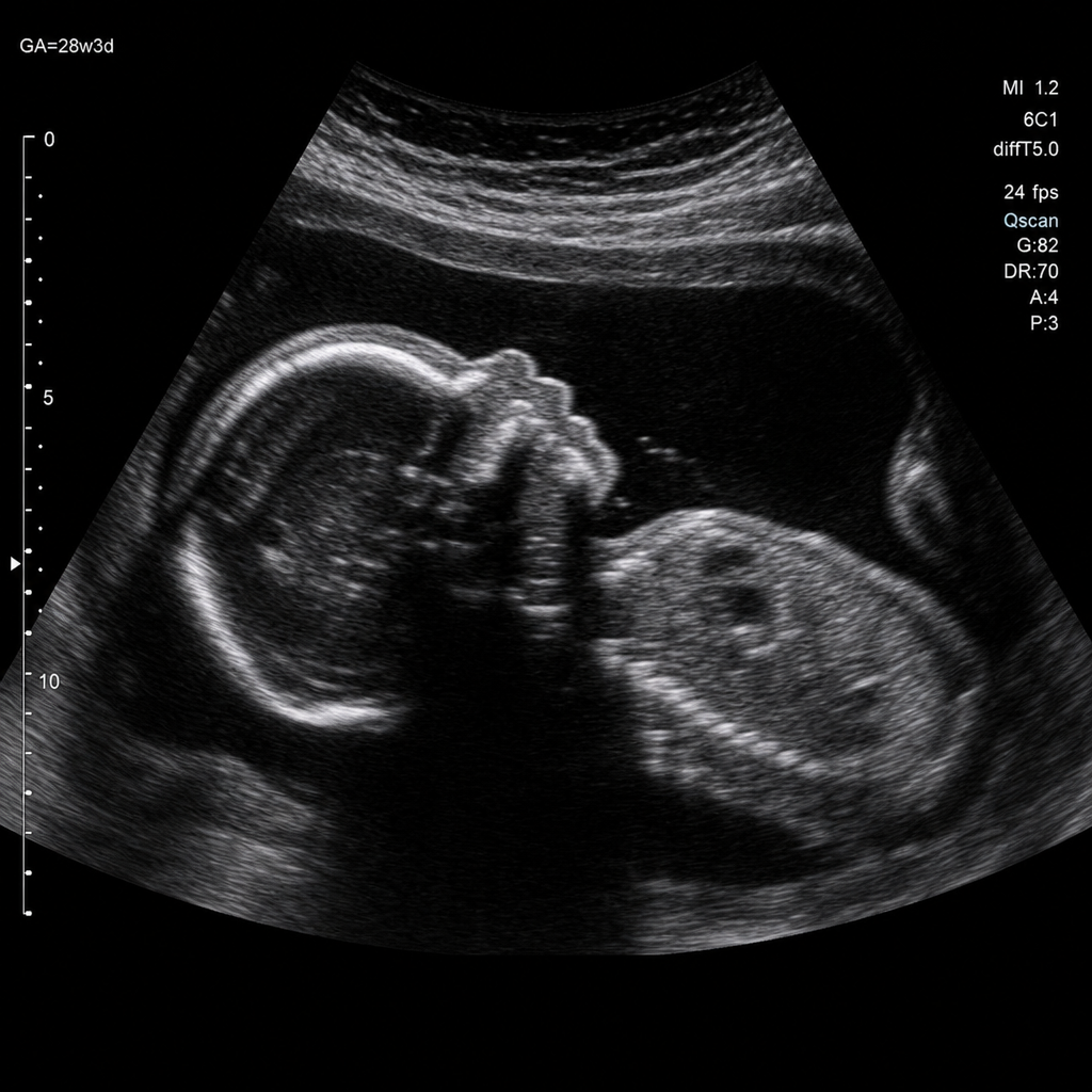
Which maternal condition is associated with the following fetal anomaly?
On a routine first-trimester ultrasound at 6 weeks, the gestational sac is seen separate from the endometrium and more than 1 cm away from the most lateral edge of the uterine cavity. The "interstitial line sign" is observed. What is your diagnosis?
Placental villi invading through the serosa of the uterus falls under which category?
The McAfee and Johnson regimen is used for which of the following conditions?
A non-stress test (NST) performed on a 36-week pregnant diabetic female is non-reactive. What is the recommended next step?
Practice by Chapter
Fetal Assessment Techniques
Practice Questions
Hypertensive Disorders in Pregnancy
Practice Questions
Intrauterine Growth Restriction
Practice Questions
Multiple Gestation
Practice Questions
Rh Isoimmunization and Other Blood Group Incompatibilities
Practice Questions
Intrauterine Fetal Therapy
Practice Questions
Prenatal Diagnosis and Genetic Counseling
Practice Questions
Placental Abnormalities
Practice Questions
Preterm Labor and Delivery
Practice Questions
Management of Medical Disorders in Pregnancy
Practice Questions
Want unlimited practice?
Get full access to all questions, explanations, and performance tracking.
Scan to download app