
Maternal-Fetal Medicine — MCQs

On this page
Mr. and Mrs. Annadurai have a 2-month-old baby with Down syndrome. Karyotype of Mrs. Annadurai shows a translocation variety of Down syndrome. Which of the following investigations will you advise to the parents before their next pregnancy?
What is the normal beat-to-beat variability in a Non-Stress Test (NST) on a term fetus?
Which of the following is NOT typically performed in the management of Intrauterine Growth Restriction (IUGR)?
A 37-week pregnant female presents with grade 3 placenta previa, vaginal bleeding, and uterine contractions. What is the most appropriate management?
A 30-year-old pregnant woman visits her obstetrician for prenatal care. The patient reports that two of her three children had "yellow jaundice" at birth, and her youngest child was severely jaundiced and required two blood transfusions. Prenatal laboratory tests reveal the mother is blood type O, Rh negative, and her husband is blood type A, Rh positive. The obstetrician obtains amniotic fluid at 36 weeks of gestation to determine if the fetus is mature enough for preterm delivery. Which of the following quantitative analyses of amniotic fluid is most likely used as an indicator of fetal lung maturity?
An HIV-infected pregnant woman presents for her 36-week prenatal visit. Which of the following discussion points regarding delivery is true?
A female has a history of 6 weeks amenorrhea, and ultrasound shows no sac. Serum beta-hCG is 1000 IU/L. What is the next step in the management of this patient?
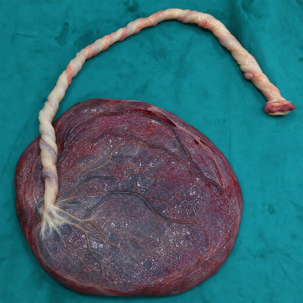
What type of placental insertion is shown?
Which of the following statements concerning polyhydramnios is true?
A multigravida with previous 2 normal deliveries presents with an unstable lie of the fetus at 34 weeks gestation. What is the most probable cause?
Practice by Chapter
Fetal Assessment Techniques
Practice Questions
Hypertensive Disorders in Pregnancy
Practice Questions
Intrauterine Growth Restriction
Practice Questions
Multiple Gestation
Practice Questions
Rh Isoimmunization and Other Blood Group Incompatibilities
Practice Questions
Intrauterine Fetal Therapy
Practice Questions
Prenatal Diagnosis and Genetic Counseling
Practice Questions
Placental Abnormalities
Practice Questions
Preterm Labor and Delivery
Practice Questions
Management of Medical Disorders in Pregnancy
Practice Questions
Want unlimited practice?
Get full access to all questions, explanations, and performance tracking.
Scan to download app