Bleeding is of fetal origin in which of the following conditions?
Which one of the following perinatal infections has the highest risk of fetal infection in the first trimester?
A 27-year-old G2P1 woman at 34 weeks' gestation presents to the emergency department following a motor vehicle collision. Her heart rate is 130/min and blood pressure is 150/90 mm Hg. She is alert and oriented to person, place, and time. She complains of severe abdominal pain that began immediately after the collision. Physical examination reveals bruising over her abdomen, along with a hypertonic uterus and dark vaginal bleeding. A sonogram reveals a placental abruption, and the fetal heart tracing reveals some decelerations. Emergency laboratory tests reveal an International Normalized Ratio of 2.5, with elevated fibrin degradation products. Which of the following is the most appropriate first step in management?
A pseudogestational sac is typically seen in which of the following conditions?
Which of the following statements is true regarding preeclampsia?
A female has a history of 6 weeks amenorrhea, ultrasonography shows an empty sac, and serum hCG is 6500 IU/L. What is the next step in management?
All are true about pre-eclampsia except:
What is the tocolytic of choice in a pregnancy with heart disease?
Which of the following is NOT considered a pregnancy morbidity associated with Antiphospholipid Antibody Syndrome (APLAS)?
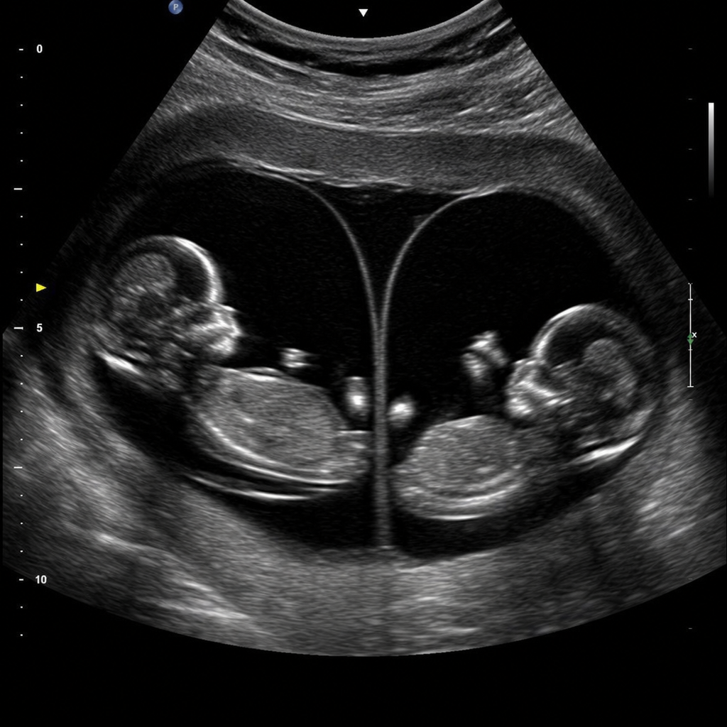
Which of the following is NOT considered a high-risk pregnancy?
Explanation: **Explanation:** In **Vasa Previa**, fetal blood vessels (unprotected by Wharton’s jelly or placental tissue) run across the internal os of the cervix, usually due to a velamentous cord insertion or succenturiate lobe. When the membranes rupture (spontaneously or artificially), these fetal vessels are lacerated. Because the blood lost comes directly from the fetal circulation, even a small amount of bleeding can lead to rapid fetal exsanguination and distress, while the mother remains hemodynamically stable. **Analysis of Incorrect Options:** * **Placenta Accreta, Increta, and Percreta:** These are spectrums of morbidly adherent placenta where the chorionic villi invade the myometrium to varying depths (Accreta: superficial; Increta: into myometrium; Percreta: through serosa). Bleeding in these conditions occurs during attempts at placental delivery and is **maternal** in origin (from the uterine spiral arteries). **High-Yield Clinical Pearls for NEET-PG:** * **The Classic Triad:** Rupture of membranes + Painless vaginal bleeding + Fetal bradycardia/distress. * **Diagnostic Test:** To differentiate fetal from maternal blood, the **Apt test** or **Ogita test** is used (based on the resistance of fetal hemoglobin to alkali denaturation). * **Antenatal Diagnosis:** Color Doppler ultrasound is the gold standard for identifying vessels crossing the os. * **Management:** If diagnosed antenatally, a planned Cesarean section is performed (usually at 34–36 weeks) to avoid labor and membrane rupture.
Explanation: **Explanation:** The risk of vertical transmission in many perinatal infections varies significantly with gestational age. For **Rubella**, the risk of fetal infection is highest during the **first trimester** (up to 80–90%). As the pregnancy progresses, the risk decreases significantly (25–30% in the second trimester) before rising again near term. More importantly, the severity of organogenesis defects (Congenital Rubella Syndrome) is maximal if the infection occurs before 12 weeks. **Analysis of Options:** * **Hepatitis B (HBV):** The risk of transmission is lowest in the first trimester (~10%) and highest in the **third trimester** (~90%). Transmission usually occurs during delivery (peripartum). * **Syphilis:** While *Treponema pallidum* can cross the placenta at any stage, the risk of transmission **increases** as pregnancy advances. It is higher in the third trimester compared to the first. * **Toxoplasmosis:** Similar to Syphilis and HBV, the rate of transmission is lowest in the first trimester (~15%) and highest in the **third trimester** (~60%). However, the severity of fetal damage is inversely proportional to the transmission risk (most severe in the first trimester). **Clinical Pearls for NEET-PG:** * **Rubella:** The "Classic Triad" of Congenital Rubella Syndrome includes **Cataracts, Sensorineural deafness, and PDA** (Patent Ductus Arteriosus). * **Rule of Inverse Relationship:** For Toxoplasmosis and Rubella, the **risk** of transmission increases with gestational age (except for Rubella's first-trimester peak), but the **severity** of fetal damage is greatest in the first trimester. * **Vaccination:** Rubella is a live-attenuated vaccine (RA 27/3 strain); it is contraindicated during pregnancy, and pregnancy should be avoided for 1 month post-vaccination.
Explanation: **Explanation:** The patient presents with classic signs of **Placental Abruption** (abdominal pain, hypertonic uterus, vaginal bleeding) following trauma. The critical finding here is the laboratory evidence of **Disseminated Intravascular Coagulation (DIC)**: an elevated INR (2.5) and increased fibrin degradation products. **1. Why "Administer fresh frozen plasma" is correct:** In the setting of placental abruption, the release of tissue thromboplastin into the maternal circulation can trigger a massive consumption of clotting factors. The patient is hemodynamically unstable (tachycardia 130/min) and has laboratory evidence of coagulopathy. **Maternal stabilization is the absolute priority** before any surgical intervention. Administering Fresh Frozen Plasma (FFP) replaces depleted clotting factors and helps correct the DIC, preventing fatal hemorrhage during or after delivery. **2. Why the other options are incorrect:** * **A. Tocolytics:** These are contraindicated in placental abruption and fetal distress. They can mask symptoms and worsen maternal hemorrhage by preventing uterine contraction. * **B. Corticosteroids:** While indicated for fetal lung maturity at 34 weeks, they take 24–48 hours to be effective. In an emergency involving DIC and fetal distress, they are not the *first* step. * **D. Deliver by LSCS:** While delivery is ultimately necessary due to fetal decelerations and abruption, performing surgery on a patient with uncorrected DIC (INR 2.5) is catastrophic and leads to uncontrollable surgical bleeding. **Stabilize the mother first.** **Clinical Pearls for NEET-PG:** * **Most common cause of DIC in pregnancy:** Abruptio Placentae. * **Management Rule:** In any obstetric emergency, "Stabilize the mother first, then address the fetus." * **Classic Triad of Abruption:** Painful vaginal bleeding, uterine tenderness/hypertonicity, and fetal distress. * **Target in DIC:** Maintain Fibrinogen >150 mg/dL and Platelets >50,000/mm³.
Explanation: ### Explanation **1. Why Ectopic Pregnancy is Correct:** A **pseudogestational sac** is a collection of fluid or blood within the uterine cavity that mimics a true gestational sac. It occurs in approximately 10–20% of ectopic pregnancies. The underlying mechanism is the hormonal stimulation of the endometrium (decidualization) by the ectopic pregnancy, leading to the breakdown of the decidua and the accumulation of fluid in the uterine lumen. **Key Distinguishing Features:** * **Location:** It is centrally located in the uterine cavity (a true sac is eccentric). * **Structure:** It lacks the "Double Decidual Sign" (the two concentric rings representing the decidua capsularis and decidua parietalis) and does not contain a yolk sac or embryo. **2. Analysis of Incorrect Options:** * **Missed Abortion:** This involves a non-viable intrauterine pregnancy. A true gestational sac is present, though it may be irregular or lack fetal cardiac activity. * **Complete Abortion:** The uterus is typically empty with a thin endometrial stripe, as all products of conception have been expelled. * **Hematometra:** This is a collection of blood in the uterus due to an anatomical obstruction (e.g., imperforate hymen). While it is a fluid collection, it does not mimic the specific appearance of a gestational sac in the context of pregnancy. **3. NEET-PG High-Yield Pearls:** * **Double Decidual Sign:** The most reliable early USG sign of an **intrauterine** pregnancy (seen at ~5.5 weeks). * **Yolk Sac:** Its presence 100% confirms an intrauterine pregnancy and rules out a pseudogestational sac. * **Discriminatory Zone:** If the serum β-hCG is >1500–2000 mIU/mL and no intrauterine sac is seen, suspect ectopic pregnancy. * **Arias-Stella Reaction:** A histological change in the endometrium (hypersecretory glands) associated with ectopic pregnancy, often found alongside a pseudogestational sac.
Explanation: **Explanation:** **1. Why the Correct Answer is Right:** The fundamental pathophysiology of preeclampsia is **systemic endothelial dysfunction**. It begins with defective trophoblastic invasion of the spiral arteries (Stage 1), leading to placental hypoxia. This triggers the release of anti-angiogenic factors (like sFlt-1 and soluble endoglin) into the maternal circulation. These factors neutralize VEGF and PlGF, causing widespread endothelial damage (Stage 2). This damage leads to increased vascular permeability (edema), activation of the coagulation cascade (thrombocytopenia), and vasospasm (hypertension and end-organ damage). **2. Why the Other Options are Incorrect:** * **Option A:** Preeclampsia is **not totally preventable**. While low-dose Aspirin (started before 16 weeks) can reduce risk in high-risk patients, the exact etiology remains multifactorial and often unpredictable. * **Option B:** Traditionally, the **diastolic blood pressure** was considered more significant as it reflects peripheral vascular resistance. However, current ACOG/NHBPEP guidelines give equal importance to both systolic (≥140 mmHg) and diastolic (≥90 mmHg) values for diagnosis. * **Option C:** Eclampsia is **not invariably preceded** by preeclampsia symptoms. In about 20-30% of cases, seizures occur without prior warning signs like significant hypertension or proteinuria (atypical eclampsia). **3. High-Yield Clinical Pearls for NEET-PG:** * **Definition:** BP ≥140/90 mmHg after 20 weeks of gestation + proteinuria OR signs of end-organ dysfunction. * **Gold Standard for Proteinuria:** Urinary protein excretion ≥300 mg in a 24-hour collection. * **Predictive Marker:** Increased sFlt-1:PlGF ratio. * **Drug of Choice (DOC):** For seizure prophylaxis/control is **Magnesium Sulfate (MgSO₄)** (Pritchard or Zuspan regimen). * **Definitive Treatment:** Delivery of the fetus and placenta.
Explanation: **Explanation:** The clinical scenario describes a case of **Early Pregnancy Loss (Anembryonic Pregnancy/Blighted Ovum)**. The diagnosis is confirmed using the concept of the **Discriminatory Zone**. 1. **Why Medical Management is correct:** The discriminatory zone is the serum hCG level above which a gestational sac should be visible on ultrasound. For Transvaginal Sonography (TVS), this is typically **1500–2000 IU/L**. In this patient, the hCG is **6500 IU/L** with an empty sac, which is well above the threshold. According to ACOG and NICE guidelines, a diagnosis of a non-viable pregnancy is made when the Mean Sac Diameter (MSD) is $\geq$ 25 mm with no embryo, or when the hCG is significantly above the discriminatory zone with no intrauterine evidence of a viable pregnancy. Once non-viability is confirmed, management options include expectant, medical (Misoprostol), or surgical (D&C). In NEET-PG contexts, for a stable patient with a confirmed non-viable pregnancy, **Medical Management** is the preferred first-line intervention. 2. **Why other options are incorrect:** * **B & C (Repeat hCG):** These are indicated in "Pregnancy of Unknown Location" (PUL) where hCG is *below* the discriminatory zone. Since the hCG here is 6500 IU/L, the diagnosis is already certain; waiting only delays treatment. * **D (Surgical Management):** While an option, it is usually reserved for patients with heavy bleeding, hemodynamic instability, or those who fail medical management. **High-Yield Clinical Pearls for NEET-PG:** * **Discriminatory Zone:** TVS = 1500–2000 IU/L; TAS (Transabdominal) = 6500 IU/L. * **Failed Pregnancy Criteria (USG):** * CRL $\geq$ 7 mm with no cardiac activity. * MSD $\geq$ 25 mm with no embryo. * **Medical Management Drug of Choice:** Misoprostol (Prostaglandin E1 analogue).
Explanation: **Explanation:** Pre-eclampsia is a multisystem disorder characterized by widespread endothelial dysfunction and vasospasm. The correct answer is **D (Deep vein thrombosis)** because, while pregnancy itself is a hypercoagulable state, DVT is not a direct pathological component or a classic complication of the pre-eclampsia disease process. **Why the other options are incorrect (Complications of Pre-eclampsia):** * **Cerebral Hemorrhage:** This is the most common cause of maternal death in eclampsia. It results from a loss of cerebrovascular autoregulation due to severe hypertension, leading to hyperperfusion and rupture of small vessels. * **Pulmonary Edema:** Occurs in about 2-5% of severe pre-eclampsia cases. It is caused by increased capillary permeability (leaky endothelium), decreased plasma oncotic pressure (due to proteinuria/hypoalbuminemia), and occasionally left ventricular failure. * **Acute Renal Failure (ARF):** Pre-eclampsia causes a characteristic renal lesion called **Glomerular Endotheliosis**. Severe vasospasm and reduced renal perfusion can lead to acute tubular necrosis (ATN) or, in extreme cases, cortical necrosis. **High-Yield Clinical Pearls for NEET-PG:** * **Definition:** BP ≥140/90 mmHg after 20 weeks of gestation with proteinuria or evidence of end-organ dysfunction. * **Pathogenesis:** Failure of secondary trophoblastic invasion of spiral arteries (high resistance remains). * **DOC for Seizure Prophylaxis:** Magnesium Sulfate ($MgSO_4$) – Pritchard or Zuspan regimen. * **Definitive Treatment:** Delivery of the fetus and placenta. * **Predictive Marker:** Low PIGF (Placental Growth Factor) and high sFlt-1 levels.
Explanation: **Explanation:** The management of preterm labor in patients with cardiac disease requires a tocolytic with the least hemodynamic impact. **Nifedipine**, a Calcium Channel Blocker (CCB), is the tocolytic of choice in this scenario. It is preferred because it is orally administered, highly effective, and has a relatively safe profile compared to other agents. While it can cause peripheral vasodilation and mild tachycardia, it lacks the severe fluid-loading requirements or significant cardiac stress associated with other classes. **Analysis of Options:** * **Nifedipine (Correct):** It is the first-line tocolytic for most pregnancies, including those with heart disease, due to its ease of use and lower risk of serious maternal cardiovascular complications. * **Magnesium Sulfate:** While used for neuroprotection, it is not the primary tocolytic of choice. In cardiac patients, there is a high risk of **pulmonary edema** and fluid overload, making it a risky option. * **Atosiban:** This oxytocin receptor antagonist is extremely safe for the heart; however, due to its high cost and limited availability in many Indian clinical settings, Nifedipine remains the standard "choice" for exams. (Note: If Nifedipine is not an option, Atosiban is the safest alternative). * **Alcohol:** Historically used, it is now obsolete due to maternal toxicity and poor efficacy. **NEET-PG High-Yield Pearls:** * **Beta-mimetics (Ritodrine/Terbutaline):** Strictly **contraindicated** in heart disease as they cause tachycardia, hypotension, and pulmonary edema. * **Indomethacin:** Avoided after 32 weeks due to the risk of premature closure of the Ductus Arteriosus. * **Drug of choice for Tocolysis (General):** Nifedipine. * **Drug of choice for Tocolysis (Diabetes):** Nifedipine (Avoid Beta-mimetics as they cause hyperglycemia).
Explanation: ### Explanation The diagnosis of **Antiphospholipid Antibody Syndrome (APLAS)** in pregnancy is based on the **Revised Sapporo Criteria (Sydney Criteria)**, which require at least one clinical criterion and one laboratory criterion (Lupus anticoagulant, Anticardiolipin, or Anti-β2 glycoprotein-I antibodies). **Why Option C is the correct answer:** The criteria specifically require **three or more** consecutive unexplained spontaneous abortions **before the 10th week** of gestation (not just "before the second trimester"). The term "second trimester" (starting at 13-14 weeks) is too broad and inaccurate according to the formal diagnostic guidelines. **Analysis of other options:** * **Option A:** One or more unexplained deaths of a morphologically normal fetus at or **beyond the 10th week** of gestation is a primary clinical criterion. * **Option B:** Premature birth **before 34 weeks** due to severe pre-eclampsia, eclampsia, or placental insufficiency is a recognized clinical criterion. * **Option D:** This option is technically incorrect in the context of the question's phrasing, as the presence of APLAS *requires* specific morbidities or vascular thrombosis to be diagnosed. ### High-Yield Clinical Pearls for NEET-PG: * **Drug of Choice:** For pregnant women with APLAS, the standard treatment is **Low Dose Aspirin (LDA) + Prophylactic Low Molecular Weight Heparin (LMWH)**. Warfarin is contraindicated due to teratogenicity. * **Laboratory Timing:** Antibodies must be present on two or more occasions, at least **12 weeks apart**. * **Mechanism:** APLAS causes a hypercoagulable state leading to placental infarction and spiral artery thrombosis. * **Most Specific Test:** Lupus Anticoagulant (LAC) is the most specific predictor of pregnancy loss among the three antibodies.
Explanation: **Explanation:** In obstetrics, a **high-risk pregnancy** is one where the mother, fetus, or newborn is at an increased risk of adverse health outcomes due to pre-existing conditions or complications arising during gestation. **Why "Third pregnancy" is the correct answer:** Parity alone does not define a high-risk pregnancy. A third pregnancy (Gravida 3, Para 2) is considered a routine obstetric case unless accompanied by specific risk factors (e.g., age >35, previous C-section, or medical comorbidities). In contrast, **Grand Multiparity** (usually defined as ≥5 previous pregnancies) is considered high-risk due to increased risks of postpartum hemorrhage (PPH), malpresentations, and placenta previa. **Analysis of Incorrect Options:** * **Rh Isoimmunization:** This is a classic high-risk condition. Maternal antibodies cross the placenta, leading to fetal hemolysis, hydrops fetalis, and intrauterine fetal death (IUFD). It requires intensive monitoring via MCA-PSV doppler and potential intrauterine transfusions. * **ABO Incompatibility:** While generally milder than Rh isoimmunization, it is a pathological state that can lead to neonatal jaundice and mild anemia, requiring maternal-fetal surveillance. * **Twin Pregnancy:** All multifetal gestations are high-risk. They carry significantly higher rates of preterm labor, pre-eclampsia, gestational diabetes, IUGR, and complications like Twin-to-Twin Transfusion Syndrome (TTTS). **NEET-PG High-Yield Pearls:** * **Elderly Primigravida:** Age >35 years at the time of first delivery is a high-risk factor. * **Short Stature:** Height <140–145 cm is a risk factor for Cephalopelvic Disproportion (CPD). * **Bad Obstetric History (BOH):** Defined as two or more consecutive spontaneous abortions, early neonatal death, or stillbirths. * **The "Too Close" Rule:** A pregnancy interval of less than 2 years is considered high-risk.
Fetal Assessment Techniques
Practice Questions
Hypertensive Disorders in Pregnancy
Practice Questions
Intrauterine Growth Restriction
Practice Questions
Multiple Gestation
Practice Questions
Rh Isoimmunization and Other Blood Group Incompatibilities
Practice Questions
Intrauterine Fetal Therapy
Practice Questions
Prenatal Diagnosis and Genetic Counseling
Practice Questions
Placental Abnormalities
Practice Questions
Preterm Labor and Delivery
Practice Questions
Management of Medical Disorders in Pregnancy
Practice Questions
Get full access to all questions, explanations, and performance tracking.
Start For Free