The normal rate of dilatation of the cervix in a primigravida in the active phase of labor is
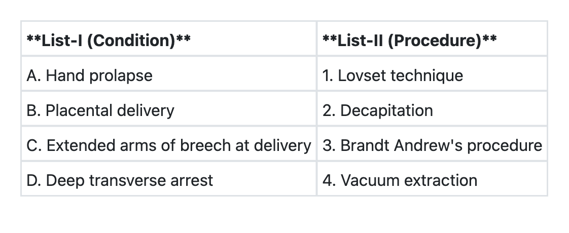
Match List-I with List-II and select the correct answer using the code given below the Lists:

A patient who just delivered at home presents with a third degree perineal tear. You will do the repair:
Face to pubes delivery occurs in which of the foetal position?
A G 2 P 1 A 0 presents with full term pregnancy with transverse lie in the first stage of labour. On examination, cervix is 5 cm dilated, membranes are intact and foetal heart sounds are regular. The appropriate management would be:
Which of the following methods for induction of labour should not be used in the patient with previous lower segment caesarean section?
Which one of the following is not a complication of shoulder dystocia?
Immediately after third stage of labour in a case of full term delivery, the fundus of the uterus is:
A 20 year old primigravida is admitted with full term pregnancy and labour pains. At 4 A.M. she goes into active phase of labour with 4 cm cervical dilatation. Membranes rupture during p/v examination showing clear liquor. A repeat p/v after 4 hours of good uterine contractions reveals a cervical dilatation of 5 cm. What should be the next step in management?
Which one of the following is not a suitable condition for outlet forceps application?
Explanation: ***1 cm/hour*** - The **active phase** of labor in a **primigravida** (first-time mother) is characterized by a cervical dilatation rate of at least 1 cm per hour. - This rate signifies good progress and is often used as a benchmark on a **partogram** to monitor labor. *0.25 cm/hour* - This rate is significantly **slower** than normal for the active phase of labor in a primigravida and would indicate **abnormal labor progression**, possibly requiring intervention. - Such a slow rate might be seen in the **latent phase** or in cases of **protracted labor**. *0.75 cm/hour* - While closer, this rate is still **below the expected minimum** for a primigravida in the active phase, suggesting slightly slower than optimal progress. - It could still indicate a **protracted active phase**, particularly if it persists. *0.5 cm/hour* - This rate is **substantially slower** than the typical progress in the active phase of labor for a primigravida. - It would be a strong indicator of **failure to progress** and would likely warrant a thorough evaluation for potential causes such as **cephalopelvic disproportion** or ineffective uterine contractions.
Explanation: ***A→2 B→3 C→1 D→4*** - **Hand prolapse (A)** occurs when a fetal hand prolapses alongside the presenting part, leading to obstructed labor. If the fetus is dead and vaginal delivery is impossible due to severe obstruction, **Decapitation (2)** may be performed as a destructive procedure to facilitate delivery. - **Placental delivery (B)** is managed by the **Brandt-Andrews maneuver (3)**, which involves controlled cord traction with counter-pressure on the uterus to prevent uterine inversion and facilitate safe placental separation. - **Extended arms of breech at delivery (C)** occurs when the fetal arms are extended above the head during breech presentation. The **Lovset maneuver (1)** is specifically designed to deliver extended arms by rotating the fetus to bring the posterior arm down and anteriorly. - **Deep transverse arrest (D)** occurs when the fetal head arrests in the transverse diameter of the pelvis. **Forceps delivery or vacuum extraction (4)** can be used with manual or instrumental rotation to deliver the fetal head. *A→4 B→1 C→3 D→2* - This incorrectly matches hand prolapse with vacuum extraction, which cannot address the obstruction caused by a prolapsed hand. It also reverses the Brandt-Andrews maneuver and Lovset technique. *A→2 B→1 C→3 D→4* - This incorrectly matches placental delivery with Lovset technique (which is for breech) and extended arms with Brandt-Andrews maneuver (which is for placental delivery). *A→4 B→3 C→1 D→2* - This incorrectly matches hand prolapse with vacuum extraction and deep transverse arrest with decapitation. Decapitation is not indicated for deep transverse arrest, which can be managed with forceps or vacuum.
Explanation: ***immediately*** - Repair of a **third-degree perineal tear** should be done **immediately** after diagnosis to minimize complications like infection, pain, and long-term functional issues. - Prompt repair helps to restore **anatomical integrity** and improve outcomes for continence and discomfort. *after 24 hours.* - Delaying the repair by 24 hours increases the risk of **infection**, **edema**, and further tissue damage, making the repair more difficult and less successful. - This delay could also lead to increased **blood loss** and patient discomfort. *after 6 weeks* - Waiting 6 weeks would allow for scar tissue formation and potential infection, making a primary repair much more challenging and possibly requiring a more complex secondary repair. - This delay would significantly increase the risk of **fecal incontinence** and other long-term complications. *after 3 months* - A three-month delay is inappropriate for a fresh perineal tear as it guarantees significant **scarring**, **fibrosis**, and high risk of **infection**. - By this time, the tear would likely have healed by secondary intention, resulting in poor anatomical and functional outcomes, often necessitating a more complicated and less effective **secondary repair**.
Explanation: ***Occipito-sacral (Occipito-posterior)*** - **"Face to pubes" delivery** is the classic mechanism in **persistent occipito-posterior (OP)** positions where the occiput is directed toward the maternal sacrum. - In this position, the fetal head delivers with **maximum extension**, and the **face passes under the pubic symphysis** (hence "face to pubes"). - The occiput sweeps over the perineum posteriorly, leading to increased perineal trauma and potential for third/fourth-degree tears. - This delivery mechanism is associated with **prolonged labor, increased back pain**, and higher rates of operative delivery. *Mentoanterior* - In **mentoanterior (MA)** face presentation, the fetal chin is anterior, and delivery occurs by **flexion after the chin passes under the symphysis**. - The mechanism involves the chin sweeping the perineum, NOT "face to pubes" delivery. - While mentoanterior can deliver vaginally, the delivery mechanism is distinctly different from occipito-posterior positions. *Brow presentation* - In **brow presentation**, the head is partially extended with the **frontal bone and anterior fontanelle presenting**. - This presents the **largest diameter (mento-vertical ~13.5 cm)** to the pelvis, making vaginal delivery virtually impossible. - Almost always requires **cesarean section** for safe delivery. *Mentoposterior* - In **mentoposterior (MP)** face presentation, the fetal chin is directed posteriorly toward the maternal sacrum. - This position **cannot deliver vaginally** as further extension of the already extended head is impossible. - Requires **rotation to mentoanterior** or cesarean section for delivery.
Explanation: ***Caesarean section*** - A **transverse lie** at term in the first stage of labor is a contraindication to vaginal delivery due to the high risk of **cord prolapse**, **uterine rupture**, and fetal distress. - **Caesarean section** is the safest mode of delivery for both mother and fetus in this scenario, as it prevents these complications. *External cephalic version* - **External cephalic version** is typically attempted in cases of **breech presentation** in the late third trimester (around 36-37 weeks) to convert it to a cephalic presentation. - It is **contraindicated** in transverse lie during active labor, especially with cervical dilation, as it has a low success rate and can lead to complications such as **placental abruption** or **cord prolapse**. *Internal podalic version* - **Internal podalic version** is a procedure typically reserved for the delivery of the second twin in a **transverse or oblique lie**, or in some cases of breech presentation. - It carries significant risks, including **uterine rupture** and fetal injury, and is generally not performed for a singleton pregnancy with cervical dilation. *Wait for spontaneous evolution and expulsion* - **Spontaneous evolution** (where the fetus rotates to a longitudinal lie) is extremely rare in a transverse lie presentation at term, especially once labor has started. - Waiting for spontaneous rotation would lead to **prolonged labor**, increased risk of **uterine rupture**, and severe fetal compromise due to obstruction.
Explanation: ***Vaginal prostaglandin gel*** - **Prostaglandins** (especially **PGE2 vaginal gel**) are **absolutely contraindicated** for induction of labor in women with a previous lower segment caesarean section (LSCS). - They cause strong, sustained uterine contractions that significantly increase the risk of **uterine rupture** in a scarred uterus. - This is the **most established and widely recognized contraindication** among induction methods for VBAC candidates. - **PGE2 vaginal preparations** have the highest documented risk of uterine rupture (1-2% or higher) in scarred uteri. *Stripping of the membrane* - This method involves separating the **chorioamniotic membranes** from the lower uterine segment, which can release natural prostaglandins. - It is generally considered **safe** in women with a previous LSCS as it causes only mild, physiologic uterine activity. - Does not directly stimulate strong, unphysiologic uterine contractions like exogenous prostaglandins. *Oxytocin drip* - **Oxytocin** can be used cautiously for induction of labor in women with a previous LSCS, with close fetal and contraction monitoring. - While it can cause strong contractions, its effect is **titratable** and can be stopped immediately if hyperstimulation occurs. - It is the **preferred pharmacological method** for induction in trial of labor after cesarean (TOLAC). - Careful dose titration and continuous monitoring make it safer than prostaglandins for this indication. *Oral prostaglandin tablet* - **Oral misoprostol** also carries significant risk of uterine hyperstimulation in patients with a scarred uterus and is generally avoided in previous LSCS. - However, vaginal prostaglandin preparations are considered the **primary contraindication** in most guidelines and examination contexts due to more extensive documentation of rupture risk and less controllable absorption. - While both prostaglandin routes are problematic, vaginal gel represents the most established contraindication for VBAC induction.
Explanation: ***Placental abruption*** - **Placental abruption** is the premature detachment of the placenta from the uterine wall, typically occurring before or during labour. It is not directly caused by the mechanical obstruction during shoulder dystocia. - Complications of shoulder dystocia are primarily related to mechanical forces exerted on the baby and mother during delivery, such as nerve injuries or uterine atony, not placental conditions. *Brachial plexus palsy* - **Brachial plexus palsy** is a common complication of shoulder dystocia, often resulting from excessive lateral traction on the fetal head or neck during delivery. - This traction can stretch or tear the nerves of the brachial plexus, leading to varying degrees of paralysis or weakness in the arm and hand. *Postpartum haemorrhage* - **Postpartum haemorrhage** is a significant risk following shoulder dystocia due to the prolonged and often traumatic nature of the delivery. - The increased manipulation and potential for uterine atony or soft tissue trauma during resolution of shoulder dystocia can predispose the mother to excessive bleeding. *Facial palsy* - **Facial palsy** can occur during shoulder dystocia if there is compression or stretching of the facial nerve (cranial nerve VII) against the maternal pelvis or other structures during delivery. - While less common than brachial plexus palsy, it can result from the abnormal forces and positions during a difficult shoulder dystocia delivery.
Explanation: ***At the level of umbilicus*** - Immediately after delivery of the placenta (third stage of labor), the **fundus** typically contracts down to the level of the **umbilicus**. - This contraction helps to compress uterine blood vessels and prevent **postpartum hemorrhage**. *At the level of xiphisternum* - The uterus reaches the **xiphisternum** only in late third trimester, as the fetus grows. - After delivery, the uterus significantly reduces in size, so it would not remain this high. *Below the level of umbilicus* - While the uterus will eventually descend below the umbilicus during **involution** (shrinking back to pre-pregnancy size), this process takes several days to weeks, not immediately after delivery. - A fundus below the umbilicus immediately after delivery might suggest a failure of proper contraction. *Just above the symphysis pubis* - The uterus is at the level of the **symphysis pubis** much earlier in pregnancy, typically around **12 weeks gestation**. - A fundus at this level immediately after delivery would indicate an abnormally small uterus or an incomplete emptying.
Explanation: ***Reassess for occipitoposterior position and cephalopelvic disproportion*** - The patient has **protracted active phase** with only 1 cm cervical dilatation in 4 hours (from 4 cm to 5 cm), which is significantly slower than the expected rate of at least 1 cm/hour in primigravidas. - Crucially, the question states she has **"good uterine contractions"**, which means the slow progress is NOT due to inadequate uterine activity. - When labor progress is slow DESPITE adequate contractions, this indicates a **mechanical obstruction** such as occipitoposterior position, cephalopelvic disproportion, asynclitism, or other malpresentation. - The next step is to **assess for these mechanical factors** through clinical examination (abdominal palpation, vaginal examination to assess position, station, molding, caput) before considering augmentation. - **Augmentation with oxytocin is contraindicated** when contractions are already adequate, as it may lead to uterine hyperstimulation without improving progress if there's mechanical obstruction. *Oxytocin drip* - Oxytocin augmentation is indicated for **hypotonic uterine dysfunction** (inadequate contractions causing slow progress). - In this case, contractions are described as **"good"**, so oxytocin is NOT appropriate as the first-line intervention. - Using oxytocin when contractions are already adequate without first ruling out mechanical obstruction can be dangerous and may lead to uterine rupture or fetal compromise. *Immediate caesarean section* - While caesarean section may ultimately be needed if mechanical obstruction is confirmed, it is **premature** without first assessing the cause of slow progress. - A diagnosis must be established before proceeding to operative delivery. *Reassess after 4 hours* - Further expectant management without intervention or diagnosis is **inappropriate** as the patient has already demonstrated inadequate progress. - Prolonged labor increases risks of maternal exhaustion, infection, and fetal compromise. - Active management requires diagnosis and intervention, not continued observation.
Explanation: ***Head is above ischial spine level*** - For **outlet forceps** application, the fetal head must be engaged, meaning the **leading point of the skull** is at or below the **level of the ischial spines (+2 station or lower)**. - If the head is above the ischial spines, it indicates a higher station, making **outlet forceps** an inappropriate and potentially dangerous choice, as it could lead to fetal or maternal injury. *Cervix fully dilated* - This is a **prerequisite** for any type of **forceps delivery**, including outlet forceps. - Performing forceps delivery with a partially dilated cervix risks severe **cervical lacerations** and other maternal complications. *Membranes absent* - This condition refers to **ruptured membranes**, which is a **necessary condition** for safe forceps application. - Intact membranes would prevent proper application of the forceps blades to the fetal head and increase the risk of **fetal scalp injury**. *Vertex presentation* - **Outlet forceps** are primarily used for **vertex presentations** (head-first), where the fetal head is oriented optimally for delivery. - Other presentations, such as **breech** or **transverse**, are **contraindications** for outlet forceps and typically require **cesarean section** or other delivery methods.
Physiology of Labor
Practice Questions
Stages of Labor and Normal Progression
Practice Questions
Fetal Monitoring Techniques
Practice Questions
Pain Management in Labor
Practice Questions
Induction and Augmentation of Labor
Practice Questions
Operative Delivery (Forceps and Vacuum)
Practice Questions
Cesarean Section: Indications and Techniques
Practice Questions
Dystocia and Abnormal Labor Patterns
Practice Questions
Obstetric Emergencies
Practice Questions
Postpartum Hemorrhage Management
Practice Questions
Get full access to all questions, explanations, and performance tracking.
Start For Free