The presence of a retraction ring at the junction of upper and lower uterine segment in labour indicates
Regarding the use of a ventouse, which one of the following statements is not correct?
A 20-year-old nulli-term primigravida is brought to the casualty with labour pains for last 24 hours and a hand prolapse. On examination, she has pulse 96/min, BP 120/80 mm Hg, and mild pallor. The abdominal examination reveals the uterine height at 32 weeks, the foetus in transverse lie and absent foetal heart sounds. On vaginal examination, the left arm of the foetus is prolapsed and the foetal ribs are palpable. The pelvis is adequate. What would be the best management option?
B-Lynch stitch is applied on the uterus for the treatment of
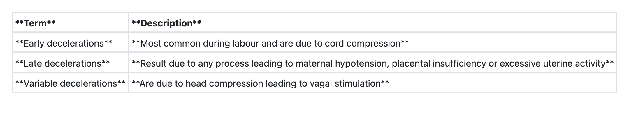
Abnormal foetal heart-rate patterns on electronic foetal monitoring include the following, except
Which one of the following is diagnosed by Spiegelberg criteria?
Regarding placental separation in III stage of labour, consider the following statements: 1. Separation of placenta occurs at decidua spongiosa 2. In Schultze method, separation of placenta starts at centre 3. In Matthews Duncan's method, separation begins at margin Which of the statements given above is/are correct?
Consider the following pairs regarding foetal heart during labour:

B-Lynch suture for atonic postpartum haemorrhage:
Which of the following genital infections is associated with preterm labour?
Explanation: ***Obstructed labour*** - A **pathological retraction ring (Bandl's ring)** forms when the upper uterine segment thickens and retracts, while the lower segment thins and distends due to **obstructed labour**. - This is distinct from the normal physiological retraction ring present in all labours—Bandl's ring is abnormally prominent, may be visible or palpable abdominally, and rises progressively higher. - This physical sign indicates an impending **uterine rupture** if the obstruction is not relieved and constitutes an obstetric emergency. *Cervical dystocia* - Refers to a cervix that fails to efface or dilate in the presence of adequate uterine contractions, but it does not directly cause the formation of a **pathological retraction ring**. - While it can lead to prolonged labour, the specific finding of a retraction ring at an abnormally high level points more directly to **obstruction**. *Precipitate labour* - Characterized by rapid labour lasting less than three hours from the onset of contractions to delivery. - It is the opposite of obstructed labour and does not involve the formation of a **pathological retraction ring**. *Prolonged labour* - Refers to labour that exceeds 20 hours for nulliparous women or 14 hours for multiparous women. - While obstructed labour can lead to prolonged labour, the presence of a **pathological retraction ring (Bandl's ring)** is a specific sign of obstruction, indicating a more severe and immediate threat than general prolongation.
Explanation: ***It can be applied when the cervix is incompletely dilated*** - A **ventouse delivery** (vacuum extraction) should only be attempted when the cervix is **fully dilated** and effaced. - Applying a ventouse to an incompletely dilated cervix risks **cervical lacerations**, uterine rupture, and significant maternal and fetal trauma. *The cup should be centrally placed on the vertex* - Proper placement of the vacuum cup is crucial for effective traction and to minimize fetal injury. - The cup should be placed over the **flexion point** (posterior fontanelle) of the fetal head, ensuring strong suction and optimal force distribution. *The largest size of the cup is preferred* - Using the **largest appropriate size** cup for vacuum extraction helps distribute the traction force over a wider area of the fetal scalp. - This reduces the risk of **scalp trauma**, such as cephalhematoma and chignon formation, by minimizing concentrated pressure. *The maximum pressure should not exceed 0.8 kg/cm²* - Maintaining the vacuum pressure below **0.8 kg/cm²** (or 50-60 cmHg) is a safety guideline to prevent excessive pressure on the fetal scalp. - Higher pressures increase the risk of **scalp lacerations**, intracranial hemorrhage, and other fetal complications.
Explanation: ***Decapitation and delivering the baby vaginally*** - With a **dead fetus in transverse lie** with **hand prolapse** and **24 hours of labor**, this represents **obstructed labor** requiring intervention. - **Decapitation** is the appropriate destructive procedure for transverse lie with shoulder presentation when the fetus is dead and vaginal delivery is feasible. - The **adequate pelvis** and **prolonged labor** (24 hours) suggest sufficient cervical dilation for vaginal delivery after decapitation. - **Foetal ribs palpable on vaginal examination** confirms adequate cervical dilation and access for the procedure. - Destructive operations are **preferred over LSCS** when the fetus is non-viable, as they avoid major abdominal surgery and its associated maternal morbidity (infection, hemorrhage, future uterine rupture risk). *Lower segment caesarean section* - While LSCS can deliver the dead fetus, it subjects the mother to **unnecessary major surgery** with higher morbidity when the fetus is already non-viable. - LSCS carries risks of **infection, hemorrhage, adhesions**, and **uterine scar complications** in future pregnancies. - When vaginal delivery is feasible after a destructive procedure, it is the preferred approach to minimize maternal trauma. *External cephalic version* - Absolutely **contraindicated** with a **dead fetus** and **hand prolapse** after 24 hours of labor. - ECV requires an **intact fetus**, adequate amniotic fluid, and is performed **before labor** or in early labor when the fetus is viable. - With established obstructed labor and fetal demise, ECV has no role. *Internal podalic version* - This procedure converts transverse or oblique lie to breech presentation to facilitate rapid vaginal delivery of a **viable second twin** or in acute situations. - It is **contraindicated** here due to **fetal demise**, **prolonged labor with potential cervical edema**, and high risk of **uterine rupture** in a primigravida with obstructed labor. - With a dead fetus, destructive procedures are safer than version and breech extraction.
Explanation: ***atonic PPH*** - The **B-Lynch stitch** is a **compression suture** applied surgically to the uterus to control severe **postpartum hemorrhage (PPH)** caused by **uterine atony**. - It works by mechanically compressing the uterus, thereby reducing blood flow and promoting uterine contraction, which is critical when the uterus fails to contract sufficiently after childbirth. *incompetent os* - An **incompetent cervical os** is typically managed with a **cervical cerclage**, a stitch placed around the cervix to prevent premature dilation during pregnancy. - The B-Lynch stitch is designed for uterine hemostasis, not cervical support. *ruptured ulcerations* - While bleeding might occur from **ruptured ulcerations**, this term is vague in an obstetrical context and does not apply to uterine bleeding specifically. - The B-Lynch stitch is used for severe uterine hemorrhage, most commonly due to atony, not general ulcerations which would require different treatment. *bleeding from placental bed of placenta previa* - **Placenta previa** bleeding often results from the placenta implanting over or near the cervix, which might require a **cesarean section** and careful placental removal. - While a B-Lynch stitch *could* be used as an adjunct in severe cases of PPH following placenta previa if atony develops, it is not the primary or typical treatment for bleeding *from the placental bed itself* which usually involves direct uterine incision or placental site hemostasis.
Explanation: ***Early decelerations*** - **Early decelerations** are considered a **benign finding** on electronic fetal monitoring, reflecting fetal head compression during contractions. - They tend to **mirror the contractions** and do not indicate fetal distress or hypoxia. *Tachycardia >170/minute lasting for 15 minutes* - Fetal **tachycardia** (heart rate >160 bpm) lasting for 10 minutes or more is considered an abnormal finding or baseline change. - A persistent fetal heart rate **>170 bpm for 15 minutes** or longer specifically indicates significant fetal tachycardia, which can be a sign of infection (e.g., chorioamnionitis), maternal fever, or fetal hypoxia. *Bradycardia <120/minute lasting for 15 minutes* - Fetal **bradycardia** (heart rate <110 bpm) lasting for 10 minutes or more is considered an abnormal finding or baseline change. - A fetal heart rate **<120 bpm lasting for 15 minutes** or longer, as specified, indicates significant fetal bradycardia, which can be associated with fetal hypoxia, cord compression, or placental insufficiency. *Late decelerations* - **Late decelerations** are a concerning sign of fetal distress, often indicative of **uteroplacental insufficiency** and fetal hypoxia. - They begin after the peak of the contraction and return to baseline after the contraction ends, reflecting a delayed fetal response to hypoxia.
Explanation: ***Ovarian pregnancy*** - Spiegelberg criteria are specifically used to diagnose an **ovarian ectopic pregnancy**, which is a rare form of ectopic pregnancy where the fertilized egg implants in the ovary. - The criteria include: the **fallopian tube and fimbria are intact** and separate from the ovary, the gestational sac is in the ovarian cortex, it is connected to the uterus by the **ovarian ligament**, and ovarian tissue can be histologically demonstrated in the sac wall. *Molar pregnancy* - This is a type of **gestational trophoblastic disease** characterized by abnormal growth of trophoblastic tissue, resulting in a non-viable pregnancy. - Diagnosis involves high levels of **hCG**, a "snowstorm" appearance on ultrasound, and histopathological examination, not Spiegelberg criteria. *Twin pregnancy* - This refers to the presence of **two fetuses** in a single pregnancy. - Diagnosis is primarily made via **ultrasound imaging** showing two distinct gestational sacs or two fetuses, and is unrelated to Spiegelberg criteria. *Uterine pregnancy* - This is a **normal intrauterine pregnancy** where the fertilized egg implants within the uterine cavity. - It is diagnosed by visualizing a gestational sac and eventually an embryo/fetus within the uterus by **ultrasound**, not by Spiegelberg criteria.
Explanation: ***1, 2 and 3*** - Correctly states that placental separation occurs at the level of the **decidua spongiosa**, which is the physiological cleavage plane permitting placental detachment after birth. - Correctly identifies that in the **Schultze method**, placental separation starts centrally, leading to the fetal surface presenting first. In the **Matthews Duncan method**, separation begins at the margin, causing the maternal surface to present first. *1 and 2 only* - This option is incorrect because while statements 1 and 2 are true, statement 3 is also correct and needs to be included for a complete answer. - It overlooks the accurate description of the **Matthews Duncan method** of placental separation. *2 and 3 only* - This option is incorrect because it fails to acknowledge the fundamental physiological fact that placental separation occurs at the **decidua spongiosa**, which is statement 1. - It omits the correct statement regarding the physiological plane of **placental separation**. *1 only* - This option is incorrect because it only includes statement 1, which is true, but excludes the correct statements 2 and 3 regarding the different methods of placental separation. - It does not account for the accurate descriptions of both the **Schultze** and **Matthews Duncan** methods.
Explanation: ***2 and 3*** - The description for **late decelerations** correctly identifies them as resulting from causes like **maternal hypotension**, **placental insufficiency**, or **excessive uterine activity**, which lead to uteroplacental insufficiency and fetal hypoxia. - The description for **variable decelerations** accurately states that they are caused by **umbilical cord compression**, which is the characteristic cause of this deceleration pattern. Variable decelerations have an abrupt onset and variable timing relative to contractions. *1 and 2* - The first statement regarding **early decelerations** is incorrect if it states they are caused by **cord compression**. Early decelerations are actually caused by **fetal head compression leading to vagal stimulation**, not cord compression. - While the second statement about late decelerations is correct, combining it with an incorrect statement about early decelerations makes this option incorrect. *2 only* - While the description for **late decelerations** is correct, this option is incomplete because the description for **variable decelerations** (statement 3) is also correct. - Answering "2 only" would imply that statement 3 is incorrect, which is not true. *1 and 3* - The first statement regarding **early decelerations** is incorrect if it attributes them to **cord compression** rather than **fetal head compression**. - While the third statement regarding **variable decelerations** is correctly described as being due to **umbilical cord compression**, the incorrectness of the first statement makes this option invalid.
Explanation: ***compresses the uterus*** - The **B-Lynch suture** is a **compression suture** applied to the uterus to mechanically reduce blood flow through sustained pressure on both anterior and posterior uterine walls. - This mechanical compression helps to achieve **haemostasis** in cases of **atonic postpartum haemorrhage** by bringing the uterine walls together and reducing the uterine cavity size. *ligates the uterine arteries* - **Uterine artery ligation** is a separate surgical procedure that involves directly tying off the uterine arteries to reduce blood flow. - The B-Lynch suture does not ligate these arteries directly; its primary mechanism is compression rather than direct vessel occlusion. *ligates the ovarian vessels* - **Ovarian artery ligation** is also a distinct surgical intervention. The B-Lynch suture is placed around the uterus and does not directly ligate the ovarian vessels. - Ovarian vessels are primarily responsible for supplying the ovaries and part of the fallopian tubes, and their ligation is not the main action of a B-Lynch suture in PPH management. *ligates the utero-ovarian anastomosis* - While there are anastomoses between the uterine and ovarian arterial systems, the B-Lynch suture does not specifically ligate these connections. - Its mechanism is general uterine compression to reduce overall blood flow and promote myometrial contraction rather than specific vessel ligation.
Explanation: ***Bacterial vaginosis*** - Bacterial vaginosis (BV) is strongly associated with an increased risk of **preterm labor** and **premature rupture of membranes** due to the production of proteases and phospholipases by anaerobic bacteria. - The imbalance of vaginal flora, particularly the overgrowth of anaerobic bacteria, can lead to ascending infection and inflammation of the **chorioamniotic membranes**. - BV has the **strongest and most consistent** evidence linking it to preterm birth among genital infections. *Human Papilloma Virus* - HPV infection is primarily known for causing **genital warts** and increasing the risk of **cervical dysplasia** and cancer. - It is not directly linked to an increased risk of preterm labor. *Monilial vaginitis* - Monilial vaginitis, or **vulvovaginal candidiasis** (yeast infection), is a common cause of vaginal discomfort, itching, and discharge. - While uncomfortable, it is not consistently associated with an increased risk of preterm labor or other adverse pregnancy outcomes. *Trichomonas vaginalis* - *Trichomonas vaginalis* infection is a sexually transmitted infection that can cause **vaginitis**, cervicitis, and urethritis. - While some studies suggest a possible association with adverse pregnancy outcomes, the evidence is **inconsistent and significantly weaker** compared to bacterial vaginosis, making BV the most established cause of preterm labor among these options.
Physiology of Labor
Practice Questions
Stages of Labor and Normal Progression
Practice Questions
Fetal Monitoring Techniques
Practice Questions
Pain Management in Labor
Practice Questions
Induction and Augmentation of Labor
Practice Questions
Operative Delivery (Forceps and Vacuum)
Practice Questions
Cesarean Section: Indications and Techniques
Practice Questions
Dystocia and Abnormal Labor Patterns
Practice Questions
Obstetric Emergencies
Practice Questions
Postpartum Hemorrhage Management
Practice Questions
Get full access to all questions, explanations, and performance tracking.
Start For Free