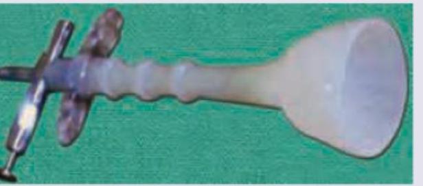
All are correct regarding the device shown here except:

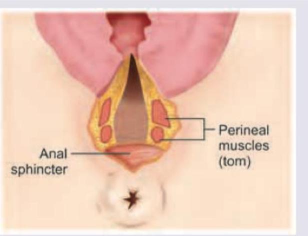
Which degree of obstetric anal sphincter injury is seen here?

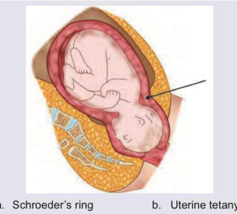
Which of the following causes of dysfunctional labour is shown below?

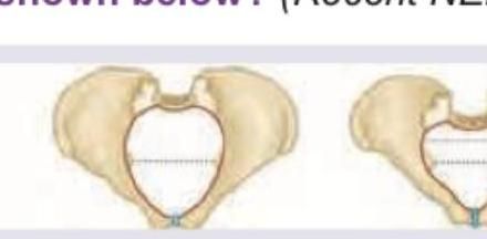
The following delivery is seen with which type of pelvis?

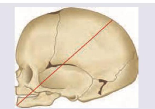
The following diameter is called \qquad and engages in \qquad presentation.

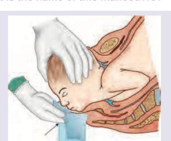
What is the name of this manoeuvre?

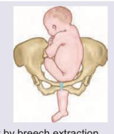
A 25-year-old primigravida at 38POG presents with painful uterine contractions and rupture of bag of water. On examination her cervix is 5 cm dilated and the malpresentation is as shown. Fetal heart rate tracing is reactive. Which of the following is the best method to achieve delivery?

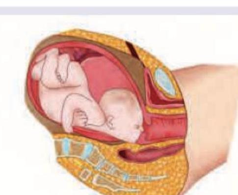
Identify the presentation shown below:

Which is the least common type of pelvis out of the types shown below?

Which is incorrect about the presentation shown below?

Explanation: ***Placed 6 cm anterior to posterior fontanelle*** - This is the **incorrect statement** - proper vacuum cup placement should be approximately **3 cm anterior to the posterior fontanelle** at the flexion point of the fetal head. - Placing the cup **6 cm anterior** would be too far forward, increasing risk of **cup dislodgement**, **subgaleal hemorrhage**, and ineffective traction. *Should generate effective vacuum of 0.8 Kg / cm^2* - This is a **correct statement** - effective vacuum pressure should be between **0.6 to 0.8 kg/cm^2** (500-600 mmHg). - This pressure range ensures adequate suction for delivery while minimizing risk of **fetal scalp injury**. *Traction at right angles to the cup* - This is a **correct statement** - traction must be applied **perpendicular to the cup plane** for optimal force transmission. - This technique prevents **cup dislodgement** and ensures efficient pulling force along the **birth canal axis**. *Traction is released in between uterine contractions* - This is a **correct statement** - traction should be **released between contractions** to prevent excessive force and allow fetal head repositioning. - Continuous traction can cause **fetal trauma** and **cup dislodgement** due to sustained pressure without uterine support.
Explanation: ***Third degree*** - The image shows a laceration involving the **anal sphincter complex** but the **anal epithelium** is intact. - This corresponds to a **third-degree** tear, where the external and/or internal anal sphincter is torn but the mucosa is preserved. *First degree* - This degree of tear involves only the **perineal skin** and **vaginal mucosa**, without affecting underlying fascia or muscle. - The image clearly shows involvement of the anal sphincter, which is beyond a first-degree tear. *Second degree* - This involves the **perineal muscles** but the **anal sphincter** is intact. - The image illustrates that the anal sphincter itself is torn, not just the perineal muscles superficial to it. *Fourth degree* - This is the most severe tear, involving the **anal sphincter complex** and extending through the **anal epithelium** (mucosa) into the lumen of the rectum. - In the depicted image, the anal epithelium appears to be intact, differentiating it from a fourth-degree injury.
Explanation: ***Bandl's ring*** - The image shows a **pathological retraction ring (Bandl's ring)**, which is an abnormal constriction of the uterus at the junction of the upper contracting segment and the lower passively dilating segment. The black arrow clearly points to the constriction. - This ring occurs when there is prolonged obstructed labor, indicating impending **uterine rupture** due to excessive thinning of the lower uterine segment. - Bandl's ring is a **sign of obstructed labor** and requires immediate intervention to prevent uterine rupture. *Uterine tetany* - **Uterine tetany** refers to excessively frequent and strong uterine contractions that are not effectively coordinated for labor progression. - This condition affects the overall uterine muscle tone and contraction pattern, rather than forming a distinct constricting ring as depicted. *Cervical dystocia* - **Cervical dystocia** is a failure of the cervix to dilate adequately, despite effective uterine contractions. - While it prolongs labor, it is a problem with cervical thinning and opening, not a physical constriction ring within the body of the uterus. *Uterine inertia* - **Uterine inertia** describes weak or infrequent uterine contractions, leading to slow or arrested labor progression. - This is characterized by a lack of effective contractile force, an entirely different issue from the pathological retraction ring shown, which indicates strong but ineffective contractions above the constriction.
Explanation: ***Anthropoid pelvis*** - This image illustrates the typical presentation of an **anthropoid pelvis**, characterized by an **oval-shaped inlet** that is wider from front to back than side to side. - This pelvic shape often facilitates a persistent **occiput posterior (OP)** position of the fetal head, as seen in the superior image, due to the longer anteroposterior diameter. *Gynecoid pelvis* - The **gynecoid pelvis** is considered the classic female pelvis, with a **rounded, slightly oval inlet** and a well-suited shape for vaginal delivery, allowing for normal fetal rotation. - It does not typically result in the persistent occiput posterior presentation and the more anterior/posterior orientation of the fetal skull shown. *Android pelvis* - An **android pelvis** has a **heart-shaped inlet** and prominent ischial spines, making it less favorable for vaginal delivery, often requiring descent in an occiput posterior position but with a narrower subpubic arch. - This type of pelvis is associated with higher rates of C-sections due to potential for cephalopelvic disproportion, and the fetal head typically encounters more difficulty at the outlet. *Platypelloid pelvis* - The **platypelloid pelvis** is characterized by a **flattened oval inlet** with a wide transverse diameter and a short anteroposterior diameter, making it the least common and least favorable for vaginal delivery. - This shape often results in the fetal head entering in a transverse arrest position, rather than the anteroposterior orientation suggested by the image.
Explanation: ***Suboccipitofrontal, Vertex*** - The diagram shows the **suboccipitofrontal diameter**, which extends from the junction of the occiput and neck to the frontal prominence (glabella). - This diameter measures approximately **10 cm** and engages in a **partially deflexed vertex presentation**. - This is one of the common engaging diameters in vertex presentations when the head is not fully flexed. *Submentobregmatic, Vertex* - The **submentobregmatic diameter** measures from the junction of the chin and neck to the bregma (anterior fontanelle). - This diameter measures approximately **9.5 cm** but engages in **face presentation, NOT vertex presentation**. - This is a critical distinction in obstetric terminology - submentobregmatic diameter is associated with face presentation. *Mentovertical, Brow* - The **mentovertical diameter** runs from the chin to the highest point of the skull (vertex). - This is the largest anteroposterior diameter of the fetal skull measuring approximately **13.5 cm**. - It is characteristic of **brow presentation**, which is often non-deliverable vaginally and may require cesarean section. *Supersubparietal, Face* - The term **supersubparietal** is not a standard fetal skull diameter used in obstetric literature. - Face presentation involves engagement of the **submentobregmatic diameter** (9.5 cm), not this non-existent diameter.
Explanation: ***Ritgen*** - The Ritgen maneuver involves applying pressure to the fetal chin through the perineum while simultaneously applying pressure to the occiput, which helps to control the **expulsion of the fetal head** and prevent perineal tears. - This maneuver assists in extending the head, guiding it through the birth canal, and protecting the maternal perineum. *Pinard* - The Pinard maneuver is used in **breech delivery** to deliver the fetal legs by abducting and flexing the thigh while exerting pressure in the popliteal fossa. - This maneuver is not applicable to a cephalic presentation, as depicted in the image. *Loveset* - The Loveset maneuver is primarily used in **breech deliveries** to rotate the fetal trunk and deliver the anterior arm first, followed by the posterior arm. - This technique is specifically for managing the arms during a breech extraction, which is not shown here. *Burns Marshall* - The Burns Marshall maneuver is another technique for delivering the aftercoming head in a **breech delivery**, where the baby's body is held in the supine position and allowed to hang, facilitating the delivery of the head by gravity and gentle traction. - This maneuver is only relevant for breech presentations and not for the cephalic presentation illustrated.
Explanation: ***Perform emergency C-section*** - The image shows a **footling breech presentation**, where one or both feet are presenting through the cervix. **Footling breech is an absolute contraindication for vaginal delivery** in modern obstetric practice. - Key risks of footling breech include **cord prolapse** (especially after rupture of membranes), incomplete cervical dilation at the time of body delivery, **entrapped aftercoming head**, and increased perinatal morbidity and mortality. - The **Term Breech Trial (Hannah et al., 2000)** established that cesarean delivery is safer than planned vaginal delivery for breech presentations at term, particularly for **footling breech** which has the worst outcomes with vaginal delivery. - Given this is a **primigravida** with **ruptured membranes** and footling breech, **emergency cesarean section is the standard of care** and the safest option for both mother and baby. *Vaginally by breech extraction* - **Vaginal breech delivery is contraindicated for footling breech** due to unacceptably high risks of cord prolapse, head entrapment, and birth trauma. - Vaginal breech delivery may only be considered in select cases of **frank or complete breech** (NOT footling), with an experienced operator, proven adequate pelvis, appropriate fetal size (2500-3800g), and flexed fetal head. - In footling breech, the small parts (feet) can pass through an incompletely dilated cervix, but the larger body and head may become entrapped, leading to catastrophic outcomes. *Perform internal podalic version* - **Internal podalic version** is a historical procedure rarely performed today, typically reserved for delivery of a **second twin in transverse lie** to convert to breech for extraction. - This procedure is **not indicated** for a singleton footling breech presentation and carries significant risks including uterine rupture, placental abruption, and fetal trauma. - The fetus is already in breech presentation; manipulating it further would not improve the situation and is contraindicated with ruptured membranes. *Deliver vaginally after external cephalic version* - **External cephalic version (ECV)** is performed **before labor** (typically 36-37 weeks) to convert breech to cephalic presentation. - ECV is **absolutely contraindicated** once the patient is in **active labor with ruptured membranes**, as attempted in this scenario at 5 cm dilation. - Attempting ECV at this stage would be ineffective, dangerous, and could cause placental abruption, cord accident, or uterine rupture.
Explanation: ***Complete breech*** - In a **complete breech** presentation, both the baby's hips and knees are **flexed**, allowing the feet to be positioned near the buttocks. - The image clearly shows the infant's knees bent and feet tucked close to the body, which is characteristic of this presentation. - This represents approximately 25% of all breech presentations. *Frank breech* - In a **frank breech** presentation, the baby's hips are flexed, but the knees are **extended**, causing the legs to be positioned straight up towards the head. - The image does not show extended knees; instead, both knees are visibly flexed. - Frank breech is the most common type, occurring in 65-70% of breech presentations. *Incomplete breech* - An **incomplete breech** (also called footling breech) occurs when one or both hips are not fully flexed, allowing one foot or knee to present below the buttocks. - This differs from the image, which shows both hips and knees fully flexed with feet tucked near the buttocks rather than presenting downward. - Incomplete breech accounts for about 10% of breech presentations. *Footling presentation* - A **footling presentation** is a type of incomplete breech where one or both of the baby's feet are positioned to deliver first. - The image clearly shows the feet tucked up near the buttocks in a flexed position rather than extended downwards or presenting first.
Explanation: ***3*** - Pelvis type 3, the **platypelloid** pelvis, is characterized by a **flattened oval inlet**, with a short anteroposterior diameter and a wide transverse diameter. - It is the **least common** type in women, representing only about **3-5%** of female pelves, and is associated with difficulties in engagement and descent of the fetal head due to the reduced anteroposterior diameter. *1* - Pelvis type 1, the **gynecoid** pelvis, is the most common and **ideal for childbirth**, found in about 50% of women. - It features a **round or slightly oval inlet**, and a wide sacrosciatic notch and subpubic angle, making it well-suited for vaginal delivery. *2* - Pelvis type 2, the **anthropoid** pelvis, has an **oval inlet** that is longer in the anteroposterior diameter than the transverse. - This type occurs in about 20-30% of women and can allow for successful vaginal delivery, often with the fetus in an occipitoposterior position. *4* - Pelvis type 4, the **android** pelvis, is characterized by a **heart-shaped inlet** and a narrow subpubic angle. - It occurs in about 20-30% of female pelves and is associated with more difficult labor due to the narrow mid-pelvis and convergent side walls.
Explanation: ***Mark commonly seen with platypelloid pelvis*** - The image shows a **brow presentation**, where the fetal head is incompletely extended, with the **forehead (bregma to root of nose)** presenting. - A **platypelloid pelvis** (flat pelvis) is characterized by a wide transverse diameter and a short anteroposterior diameter, which typically **favors transverse lie or occipito-transverse positions**, NOT brow presentation. - Brow presentation is more commonly associated with **cephalopelvic disproportion, anencephaly, multiparity, prematurity**, and other factors that prevent proper head flexion. *Associated with anencephaly* - This statement is **correct**. Brow presentation is indeed more common in fetuses with **anencephaly** due to the absence of the cranial vault, which alters fetal head mechanics and prevents normal flexion. - The altered head shape and lack of cranial vault prevent proper flexion, leading to the forehead becoming the presenting part. *Head is partially extended* - This statement is **correct**. In a **brow presentation**, the fetal head is in a state of **partial or incomplete extension**, positioned between full flexion (vertex) and full extension (face presentation). - The presenting diameter is the **mento-vertical diameter** (approximately 13-13.5 cm), which is the largest diameter and often leads to cephalopelvic disproportion. - If the head were fully extended, it would be a **face presentation** (mentum presenting); if fully flexed, it would be a **vertex presentation** (occiput presenting). *Vaginal delivery is possible* - This statement is **correct**. While **brow presentations** often lead to **cephalopelvic disproportion** and commonly require Cesarean section, vaginal delivery is possible in certain circumstances: - If the head **flexes** to a vertex presentation during labor - If the head **extends** to a face presentation during labor - If the fetus is small and the pelvis is adequately capacious - However, **persistent brow presentations** are associated with significantly increased risk of obstructed labor, maternal trauma, and perinatal morbidity, usually necessitating operative delivery.
Physiology of Labor
Practice Questions
Stages of Labor and Normal Progression
Practice Questions
Fetal Monitoring Techniques
Practice Questions
Pain Management in Labor
Practice Questions
Induction and Augmentation of Labor
Practice Questions
Operative Delivery (Forceps and Vacuum)
Practice Questions
Cesarean Section: Indications and Techniques
Practice Questions
Dystocia and Abnormal Labor Patterns
Practice Questions
Obstetric Emergencies
Practice Questions
Postpartum Hemorrhage Management
Practice Questions
Get full access to all questions, explanations, and performance tracking.
Start For Free