Which of the following is NOT related to blood coagulation disorders in obstetrics?
Saffron colored meconium is seen in which condition?
Combination of Nifedipine with what other tocolytic agent can potentially cause dangerous neuromuscular blockade?
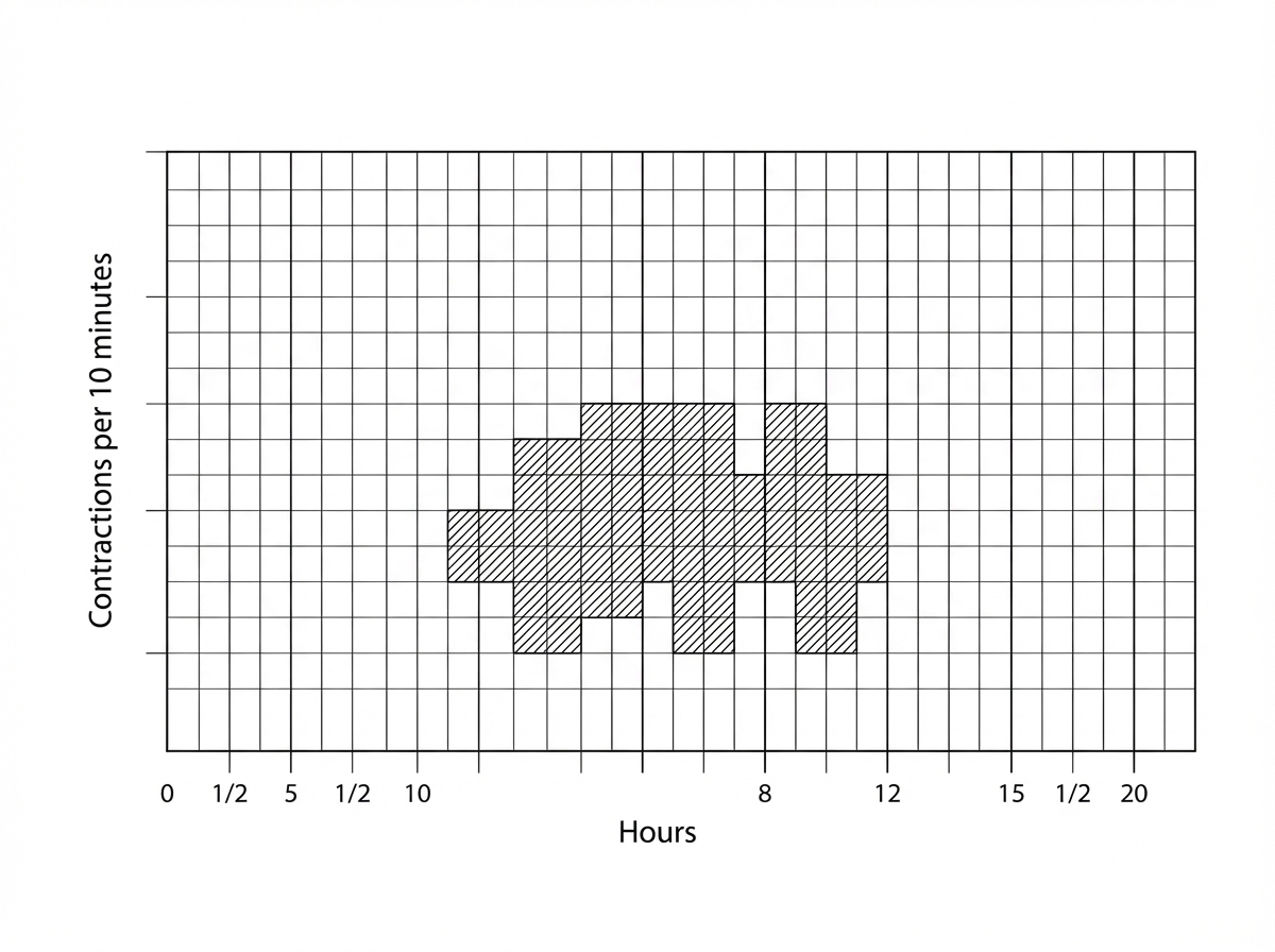
What does the indicator in the following partograph signify?

An early deceleration pattern on cardiotocography indicates what?
Pain in early labor is transmitted through which spinal nerve segments?
Prolonged latent phase of labor is commonly seen in which of the following conditions?
In twin pregnancy, vaginal delivery is contraindicated in which of the following conditions?
Antepartum hemorrhage occurs after how many weeks of gestation?
Prelabour pains are mediated through which spinal nerve roots?
Explanation: **Explanation** In the context of obstetric emergencies like Abruptio Placentae or Amniotic Fluid Embolism, distinguishing between **Disseminated Intravascular Coagulation (DIC)** and **Primary Fibrinolysis** is crucial. **Why Option A is the Correct Answer (The False Statement):** Thrombocytopenia (low platelet count) is a hallmark feature of **DIC**, not primary fibrinolysis. In DIC, there is widespread activation of the coagulation cascade leading to the "consumption" of platelets and clotting factors. In contrast, primary fibrinolysis involves the breakdown of fibrinogen/fibrin by plasmin without the initial formation of extensive microthrombi; therefore, platelets are generally not consumed and remain within normal limits. **Analysis of Other Options:** * **Option B:** In DIC, microangiopathic hemolytic anemia (MAHA) occurs as RBCs are sheared while passing through fibrin mesh in small vessels, creating **schistocytes** (helmet cells). In primary fibrinolysis, there is no fibrin meshwork to fragment the cells, so morphology remains normal. * **Option C:** The **Weiner Clot Observation Test** (Bedside Clot Test) is a high-yield clinical tool. If a clot fails to form within 6–10 minutes or dissolves rapidly, it indicates a fibrinogen level <150 mg/dL. * **Option D:** A peripheral blood smear is a rapid, reliable method to visualize a decrease in platelet density and confirm thrombocytopenia. **High-Yield Clinical Pearls for NEET-PG:** * **Most common cause of DIC in pregnancy:** Abruptio Placentae. * **Earliest lab indicator of DIC:** Increased FDPs (Fibrin Degradation Products) and D-dimer. * **Management Priority:** Always treat the underlying cause (e.g., evacuate the uterus) and replace blood components (FFP for factors, Cryoprecipitate for fibrinogen).
Explanation: **Explanation:** The color of the amniotic fluid (liquor amnii) serves as a vital clinical indicator of fetal well-being. In a normal pregnancy, liquor is clear or straw-colored. **Why Postmaturity is correct:** In **post-term pregnancies** (gestation >42 weeks), the presence of meconium is common. **Saffron-colored** (yellowish-green) meconium is a classic sign of postmaturity. This specific hue occurs because the meconium has been present in the amniotic fluid for a prolonged period, leading to the staining of the fetal skin, nails, and umbilical cord. **Analysis of Incorrect Options:** * **Toxemia of Pregnancy (Preeclampsia):** This is typically associated with **Golden-yellow** liquor, which is also seen in Rh-isoimmunization due to excessive bilirubin. * **Breech Presentation:** While meconium passage is common in breech delivery due to physical compression of the fetal abdomen during labor, it usually appears as **fresh green** meconium rather than saffron-colored. * **Normal Appearance:** Normal liquor is **clear/colorless** at term. **High-Yield Clinical Pearls for NEET-PG:** * **Green (Fresh Meconium):** Indicates acute fetal distress. * **Golden Yellow:** Rh-isoimmunization (due to bilirubin). * **Greenish-Yellow (Saffron):** Postmaturity. * **Dark Red (Corn-syrup/Meat-wash):** Accidental hemorrhage (Abruptio Placentae). * **Dark Brown (Tobacco juice):** Intrauterine Fetal Death (IUD). Understanding these color variations is essential for diagnosing underlying fetal conditions during the intrapartum period.
Explanation: ### Explanation The correct answer is **Magnesium sulfate (MgSO₄)**. **Mechanism of Interaction:** Both Nifedipine (a Calcium Channel Blocker) and Magnesium sulfate act by inhibiting calcium-related processes, but at different sites. Nifedipine blocks the entry of extracellular calcium into the myometrial cells through L-type channels. Magnesium sulfate competes with calcium at the neuromuscular junction and inhibits the release of acetylcholine. When used together, they exert a synergistic effect that can lead to profound muscle relaxation, resulting in **dangerous neuromuscular blockade**, respiratory depression, and cardiac toxicity. **Analysis of Incorrect Options:** * **A. Terbutaline:** This is a $\beta_2$-agonist. While combining it with Nifedipine can increase the risk of maternal pulmonary edema and tachycardia, it does not cause neuromuscular blockade. * **C. Indomethacin:** A COX inhibitor used as a second-line tocolytic (especially before 32 weeks). Its primary side effects are fetal (premature closure of ductus arteriosus and oligohydramnios), not neuromuscular. * **D. Atosiban:** An oxytocin receptor antagonist. It has a very specific site of action and a superior safety profile; it does not interact with Nifedipine to cause muscle paralysis. **High-Yield Clinical Pearls for NEET-PG:** * **First-line Tocolytic:** Nifedipine is currently the preferred first-line tocolytic for preterm labor (32–34 weeks) due to its oral administration and better side-effect profile. * **Antidote for MgSO₄ Toxicity:** Calcium gluconate (10 ml of 10% solution IV over 10 minutes). * **Contraindication:** Nifedipine should be avoided in patients with hypotension or cardiac disease. * **Monitoring:** When a patient is on MgSO₄, always monitor the **patellar reflex** (first sign of toxicity to disappear), respiratory rate (>12/min), and urine output (>30ml/hr).
Explanation: ***Moderate uterine contractions*** - **Hatched/lined shading** on the partograph indicates moderate uterine contractions lasting **20-40 seconds** in duration. - This represents the **standard method** for documenting contraction intensity using visual indicators on labor monitoring charts. *Mild uterine contractions* - Represented by **dotted shading** on the partograph, indicating contractions lasting **less than 20 seconds**. - These are typically **weaker contractions** that occur early in labor or during latent phase. *Strong uterine contractions* - Indicated by **solid black shading** on the partograph for contractions lasting **more than 40 seconds**. - These represent **intense contractions** typically seen during active labor and transition phases. *Arrest of labor* - This is a **clinical diagnosis** based on lack of cervical dilation or fetal descent, not a partograph shading indicator. - Would be identified by **plotting cervical dilation** crossing the action line, not by contraction shading patterns.
Explanation: **Explanation:** **Early decelerations** are characterized by a gradual decrease and return to baseline of the fetal heart rate (FHR) that is perfectly synchronous with the uterine contraction. The "nadir" (lowest point) of the deceleration occurs at the same time as the "peak" of the contraction. **Why Head Compression is Correct:** This pattern is a physiological response caused by **fetal head compression** during labor. When the head is compressed in the birth canal, it leads to increased intracranial pressure, which stimulates the **vagus nerve**. This vagal stimulation causes a transient slowing of the heart rate. Because it is a mechanical reflex rather than a sign of oxygen deprivation, early decelerations are considered **benign** and do not require intervention. **Why Other Options are Incorrect:** * **B & D (Fetal Distress/Hypoxia):** These are typically associated with **Late Decelerations** (nadir occurs after the peak of contraction), which indicate uteroplacental insufficiency and reduced fetal oxygenation. * **C (Cord Prolapse):** This usually results in **Variable Decelerations** (abrupt drop, V or U-shaped) due to umbilical cord compression, or prolonged bradycardia if the compression is sustained. **High-Yield Clinical Pearls for NEET-PG:** * **Mnemonic (VEAL CHOP):** * **V**ariable — **C**ord compression * **E**arly — **H**ead compression * **A**ccelerations — **O**kay (Fetal well-being) * **L**ate — **P**lacental insufficiency * Early decelerations are often seen during the **active stage of labor** (between 4–7 cm dilation) and during the **second stage** (pushing). * No medical management (like oxygen or C-section) is needed for isolated early decelerations.
Explanation: ### Explanation **1. Why T11 – T12 is Correct:** Pain in the **first stage of labor** (early labor) is primarily **visceral** in nature. It originates from the stretching and dilatation of the cervix and the lower uterine segment, as well as uterine contractions. These visceral afferent impulses travel alongside sympathetic nerves and enter the spinal cord through the **T10, T11, T12, and L1** nerve roots. Among the given options, T11 – T12 represents the primary segments involved in this stage. **2. Why the Other Options are Incorrect:** * **L2 – L3 & L4 – L5:** These lumbar segments do not receive primary sensory input from the uterus or the birth canal during labor. While some referred pain may be felt in the back, they are not the primary dermatomal pathways for labor pain. * **S2 – S4 (Option D):** These segments are responsible for **somatic pain** during the **second stage of labor**. This pain arises from the stretching of the vagina, perineum, and pelvic floor as the fetus descends. This is transmitted via the **pudendal nerve**. **3. Clinical Pearls for NEET-PG:** * **First Stage of Labor:** Pain is visceral, transmitted via **T10 – L1**. It is often described as dull, aching, and poorly localized. * **Second Stage of Labor:** Pain is somatic, transmitted via **S2 – S4** (Pudendal nerve). It is sharp and well-localized to the perineum. * **Regional Anesthesia:** * For a **Paracervical block**, the target is the uterine plexus (relief for the 1st stage). * For a **Pudendal block**, the target is the S2–S4 roots (relief for the 2nd stage/episiotomy). * **Epidural Analgesia** typically aims for a block from T10 to S5 to cover both stages of labor effectively.
Explanation: The **Latent Phase** of labor begins with the onset of regular uterine contractions and ends when the cervix reaches 4–6 cm dilation. According to Friedman’s criteria, a prolonged latent phase is defined as **>20 hours in primigravida** and **>14 hours in multigravida**. ### Why "Unripe Cervix" is Correct: The primary physiological goal of the latent phase is **cervical effacement** and softening. An **unripe cervix** (low Bishop score) presents high resistance to uterine contractions. This lack of readiness necessitates a longer period of contractions to achieve the structural changes required for active dilation, making it the most common cause of a prolonged latent phase. ### Analysis of Incorrect Options: * **Excessive Sedation:** While early administration of analgesics or sedatives can slow down labor, it is a secondary factor. An unripe cervix remains the most frequent clinical finding associated with this delay. * **Placenta Previa:** This is a condition where the placenta covers the internal os. It is a contraindication to vaginal delivery and typically presents with painless antepartum hemorrhage, not a delay in the latent phase. * **Abruptio Placenta:** This involves premature separation of the placenta. It often leads to uterine hypertonicity and rapid (precipitate) labor or fetal distress, rather than a prolonged latent phase. ### High-Yield NEET-PG Pearls: * **Management:** The preferred management for a prolonged latent phase is **therapeutic rest** (e.g., Morphine) or **oxytocin augmentation**. It is *not* an indication for Cesarean section. * **Friedman vs. WHO:** While Friedman used 4 cm as the start of the active phase, the **WHO Labor Care Guide (2020)** now defines the active phase starting at **5 cm** dilation. * **Most common cause overall:** Unripe cervix at the onset of labor.
Explanation: **Explanation:** The mode of delivery in twin pregnancies depends primarily on the presentation of the twins and the type of placentation. **Why Option A is Correct:** **Monochorionic Monoamniotic (MCMA) twins** are a high-risk category where both fetuses share a single amniotic sac. Vaginal delivery is **contraindicated** (elective Cesarean section is recommended at 32–34 weeks) due to the extremely high risk of **cord entanglement** and cord prolapse. As the first twin descends, the entangled cords can tighten, leading to acute fetal distress or sudden fetal demise of one or both twins. **Why the other options are Incorrect:** * **B. First twin vertex, second twin breech:** This is the most common "discordant" presentation. Vaginal delivery is generally favorable. After the birth of the first twin, the second twin can be delivered via assisted breech delivery or internal podalic version. * **C. Second twin transverse:** If the first twin is vertex, a transverse second twin is not an absolute contraindication to vaginal delivery. After the first birth, the second twin often converts to longitudinal lie spontaneously, or the clinician can perform external or internal version. * **D. First twin mentoanterior:** While a mentoposterior presentation requires a C-section, a **mentoanterior** presentation (face presentation) is a vaginal delivery-possible position, provided the pelvis is adequate and there is no cephalopelvic disproportion. **High-Yield Clinical Pearls for NEET-PG:** * **Locked Twins:** Occurs most commonly when the first twin is breech and the second is vertex (Breech-Vertex). This is a classic indication for C-section. * **Ideal Delivery Timing:** MCMA twins (32–34 weeks via C-section); MCDA twins (36–37 weeks); DCDA twins (37–38 weeks). * **Prerequisite for Vaginal Delivery:** The first twin **must** be in vertex presentation. If the first twin is non-vertex, C-section is mandatory.
Explanation: ### Explanation **1. Understanding the Correct Answer (D: 28 weeks)** Antepartum Hemorrhage (APH) is clinically defined as bleeding from or into the genital tract occurring **after the 28th week of pregnancy** but before the birth of the baby. The 28-week threshold is historically significant in the Indian context as it marks the traditional age of **fetal viability**. Bleeding during this period is most commonly associated with placental causes like Placenta Previa or Abruptio Placentae. **2. Why Other Options are Incorrect** * **A, B, and C (12, 18, and 20 weeks):** Bleeding occurring before the 28th week of gestation is not classified as APH. Instead, it is categorized under **"Bleeding in Early Pregnancy."** The most common causes for bleeding in these stages include various types of Abortions (Miscarriage), Ectopic Pregnancy, or Hydatidiform Mole. While the international definition of viability (WHO) has shifted toward 20–24 weeks in developed countries, for the purpose of standard Indian medical examinations and textbooks (like DC Dutta), 28 weeks remains the definitive cutoff for APH. **3. High-Yield Clinical Pearls for NEET-PG** * **Incidence:** APH occurs in roughly 2–5% of all pregnancies. * **Classification:** * **Placental site (70%):** Placenta Previa (painless, bright red) and Abruptio Placentae (painful, dark red). * **Extra-placental (5%):** Cervical polyps, carcinoma of the cervix, or local trauma. * **Unexplained (25%):** Where no definitive cause is found. * **Management Rule:** Any patient presenting with APH must undergo a **per-speculum examination** only after Placenta Previa is ruled out by ultrasound. A **per-vaginal (digital) examination is strictly contraindicated** until then, as it can provoke torrential hemorrhage.
Explanation: **Explanation:** The pain experienced during the first stage of labor (including prelabour or "false" pains) is primarily **visceral** in nature. It originates from the stretching of the lower uterine segment and the dilation of the cervix. 1. **Why T10–T12 is correct:** The sensory nerve fibers from the body of the uterus and the cervix travel alongside sympathetic nerves. They pass through the hypogastric plexus and enter the spinal cord via the **T10, T11, and T12** (and occasionally L1) spinal nerve roots. Therefore, pain during the early stages of labor is referred to the dermatomes supplied by these segments (the lower abdomen and lumbar region). 2. **Analysis of Incorrect Options:** * **T12–L3:** While there is some overlap at T12 and L1, the primary sensory input for uterine contractions does not extend down to L3. * **S2–S4 (Option C/S1-S3):** These roots mediate **somatic pain** during the **second stage of labor**. This pain is caused by the stretching of the pelvic floor, vagina, and perineum as the fetus descends, transmitted via the **pudendal nerve**. * **L3–L4:** These segments are generally not involved in the primary pain pathways for labor; they are more relevant for lower limb motor and sensory functions. **High-Yield Clinical Pearls for NEET-PG:** * **First Stage of Labor:** Pain is visceral, mediated by **T10–L1**. * **Second Stage of Labor:** Pain is both visceral (uterus) and somatic (perineum), mediated by **T10–L1 AND S2–S4**. * **Epidural Anesthesia:** To provide effective relief during the first stage, the block must reach the **T10** level. * **Paracervical Block:** Targets the Frankenhäuser’s plexus to relieve pain from cervical dilation (Stage 1) but does not relieve perineal pain (Stage 2).
Physiology of Labor
Practice Questions
Stages of Labor and Normal Progression
Practice Questions
Fetal Monitoring Techniques
Practice Questions
Pain Management in Labor
Practice Questions
Induction and Augmentation of Labor
Practice Questions
Operative Delivery (Forceps and Vacuum)
Practice Questions
Cesarean Section: Indications and Techniques
Practice Questions
Dystocia and Abnormal Labor Patterns
Practice Questions
Obstetric Emergencies
Practice Questions
Postpartum Hemorrhage Management
Practice Questions
Get full access to all questions, explanations, and performance tracking.
Start For Free