A pregnant patient presents with abdominal pain and bleeding per vaginam with a twin gestation of 34 weeks. What is the most probable diagnosis?
In a primigravida, when does the fetal head typically engage?
What is the best method for induction of labor in cases of hydramnios?
What is the most common cause of perinatal mortality in twins?
Episiotomy is best performed in which direction?
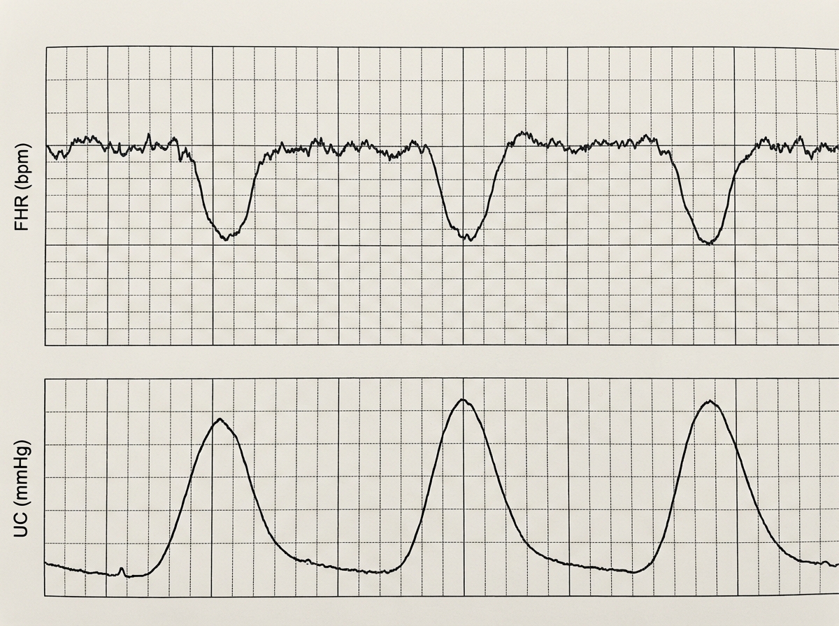
A 30-year-old female patient in labor has the following fetal tocographic finding. What does it suggest?

What is the most common breech presentation in a nulliparous woman?
Allowing the cord blood to pass to the fetus before clamping the umbilical cord should be avoided to prevent which of the following?
During delivery, the baby's head was delivered but the shoulders were not delivered after one minute. What is the next step?
A 26-year-old G2P1 woman at 41 weeks gestation has been pushing for 3 hours without progress. Her vaginal examination shows complete dilation, complete effacement, and 0 station, with the fetal head persistently in the occiput posterior position. Which of the following statements accurately describes the situation?
Explanation: **Explanation:** The clinical presentation of **abdominal pain** combined with **bleeding per vaginam** in the third trimester (34 weeks) is a classic triad for **Abruptio Placenta**. In this condition, the placenta prematurely separates from the uterine wall before delivery. **Twin gestation** is a significant risk factor for abruption due to sudden uterine decompression or chronic overdistension. The pain is typically constant and associated with uterine tenderness or hypertonicity. **Why other options are incorrect:** * **Placenta Previa:** While it presents with third-trimester bleeding, it is characteristically **painless, causeless, and recurrent**. The presence of abdominal pain strongly points away from previa toward abruption. * **Ectopic Pregnancy:** This typically presents in the **first trimester** (usually before 12 weeks) with a triad of amenorrhea, pain, and bleeding. It is not a viable diagnosis at 34 weeks. * **Abortion:** By definition, abortion refers to the termination of pregnancy **before 20–24 weeks** (depending on local guidelines). At 34 weeks, the condition is classified under Antepartum Hemorrhage (APH). **Clinical Pearls for NEET-PG:** * **Risk Factors for Abruption:** Hypertension (most common), trauma, cocaine use, polyhydramnios, and multiple gestations. * **Couvelaire Uterus:** A complication of severe abruption where blood intravasates into the myometrium, giving it a port-wine appearance. * **Coagulation Profile:** Abruptio placenta is the most common cause of **DIC** (Disseminated Intravascular Coagulation) in obstetrics. * **Management:** If the fetus is alive and in distress, immediate Cesarean section is the treatment of choice.
Explanation: ### Explanation **1. Why 36 weeks is correct:** In a primigravida (a woman pregnant for the first time), the abdominal muscles are typically firm and the lower uterine segment is well-formed. This creates sufficient pressure to push the fetal head into the pelvic brim well before the onset of labor. Engagement—defined as the passage of the widest transverse diameter of the fetal head (biparietal diameter) through the pelvic inlet—typically occurs around **36 weeks of gestation**. This phenomenon is clinically associated with "lightening," where the mother feels relief from respiratory pressure as the fetus descends. **2. Why the other options are incorrect:** * **B & D (During Labor):** In primigravidae, if the head is not engaged by the onset of labor, it is often considered a sign of cephalopelvic disproportion (CPD). Conversely, in **multigravidae**, the abdominal muscles are laxer, allowing the head to remain high and engage only after labor has commenced (1st stage). * **C (At Term):** While 37–40 weeks is "at term," the physiological standard for engagement in a first pregnancy is specifically 36 weeks. Waiting until 40 weeks to see engagement in a primigravida would be clinically late. **3. NEET-PG High-Yield Pearls:** * **Engagement Rule:** Primigravida = 36 weeks; Multigravida = During labor. * **Clinical Sign:** Engagement is confirmed on abdominal palpation when the head is **2/5ths or less** palpable above the symphysis pubis. * **Station:** On vaginal examination, engagement corresponds to the leading bony part of the fetal head reaching the level of the **ischial spines (Station 0)**. * **Non-engagement at 38 weeks** in a primigravida is a "red flag" and requires evaluation for CPD, placenta previa, or fetal anomalies.
Explanation: In cases of **Polyhydramnios (Hydramnios)**, the primary risk during induction of labor is the sudden, uncontrolled release of a large volume of amniotic fluid. This can lead to two life-threatening complications: **Cord Prolapse** (due to the fluid gush carrying the cord down) and **Abruptio Placentae** (due to the sudden decompression and shrinking of the uterine surface area). ### Why Option C is Correct **Abdominal amniocentesis followed by a stabilizing oxytocin drip** is the preferred method because it allows for a **slow, controlled decompression** of the amniotic sac. By removing fluid transabdominally before labor begins, the intrauterine pressure is reduced gradually. This stabilizes the fetal lie, prevents the sudden "snap" of the placenta, and minimizes the risk of the cord being washed down. Once the volume is reduced, an oxytocin drip is started to initiate effective uterine contractions. ### Why Other Options are Incorrect * **A & B (High/Low Rupture of Membranes):** Any form of Artificial Rupture of Membranes (ARM) in a tense hydramnios uterus carries a high risk of sudden decompression. Even a "high" rupture can lead to an uncontrollable gush, increasing the risk of cord prolapse and placental abruption. * **D (Prostaglandins):** While prostaglandins are used for cervical ripening, they do not address the mechanical issue of excessive fluid volume. Inducing contractions in a severely overdistended uterus can be ineffective or lead to uterine inertia. ### NEET-PG High-Yield Pearls * **Definition:** Polyhydramnios is defined as an Amniotic Fluid Index (AFI) > 25 cm or a Single Deepest Pocket (SDP) > 8 cm. * **Most Common Cause:** Idiopathic (60%), followed by Maternal Diabetes. * **Complication to watch for:** Postpartum Hemorrhage (PPH) due to uterine atony from overdistension. * **Management Tip:** If performing ARM in hydramnios, it should be done using a "controlled" technique (e.g., using a needle) to ensure slow fluid release.
Explanation: **Explanation:** **Prematurity (Option C)** is the single most common cause of perinatal morbidity and mortality in twin gestations. Approximately 50–60% of twin pregnancies result in preterm birth (before 37 weeks), compared to only 10% in singletons. The primary driver is uterine overdistension, which triggers early labor or premature rupture of membranes (PROM). Complications arising from prematurity—such as Respiratory Distress Syndrome (RDS), intraventricular hemorrhage, and necrotizing enterocolitis—account for the majority of neonatal deaths. **Analysis of Incorrect Options:** * **Single fetal demise (Option A):** While it increases the risk of neurological damage or death in the surviving twin (especially in monochorionic twins), it is statistically less common than complications from early delivery. * **Twin-to-Twin Transfusion Syndrome (TTTS) (Option B):** This is a serious complication unique to monochorionic diamniotic (MCDA) twins. While it has a high mortality rate if untreated, it only affects about 10–15% of monochorionic pregnancies, making it less frequent than prematurity overall. * **Intrauterine Growth Restriction (IUGR) (Option D):** Twins are at higher risk for growth restriction due to placental insufficiency or unequal sharing. While IUGR increases vulnerability, prematurity remains the leading cause of death. **High-Yield Clinical Pearls for NEET-PG:** * **Average duration of pregnancy:** Singletons (40 weeks), Twins (37 weeks), Triplets (33 weeks). * **Most common complication of twins:** Prematurity. * **Most common type of twins:** Dizygotic (70-80%). * **Vanishing Twin Syndrome:** Death of one fetus in the first trimester (occurs in ~20% of twin pregnancies). * **Bed rest:** Routine hospitalization or bed rest does *not* prevent preterm labor in twins and is not recommended.
Explanation: **Explanation:** Episiotomy is a surgically planned incision on the perineum during the second stage of labor. The **Mediolateral** approach is the gold standard and the most commonly performed technique worldwide. **1. Why Mediolateral is the Correct Answer:** The incision begins at the midpoint of the fourchette and is directed downwards and outwards at an angle of **45 degrees** toward the ischial tuberosity (usually to the right). This direction provides the best balance between increasing the vaginal outlet and protecting the anal sphincter. By directing the incision away from the midline, it significantly reduces the risk of **Third and Fourth-degree perineal tears** (extension into the anal sphincter and rectal mucosa). **2. Why Other Options are Incorrect:** * **Medial (Midline):** While it heals faster and has less blood loss, it carries a very high risk of extending into the anus, leading to rectovaginal fistulas or fecal incontinence. * **Lateral:** This incision starts 1 cm away from the midline. It is avoided because it may damage the **Bartholin’s duct** and provides poor anatomical repair. * **J-shaped:** This starts in the midline and curves laterally like a 'J' to avoid the anus. It is technically difficult to perform and repair, offering no superior benefit over the mediolateral approach. **High-Yield Clinical Pearls for NEET-PG:** * **Timing:** It should be performed when the perineum is bulging and **3-4 cm of the fetal scalp** is visible during a contraction (crowning). * **Structures Cut:** Skin, subcutaneous tissue, vaginal mucosa, **Bulbospongiosus**, and **Superficial transverse perineal muscles**. (Note: The Levator ani is usually not involved in a standard episiotomy). * **Nerve Block:** Usually performed under local infiltration or **Pudendal nerve block** (S2, S3, S4). * **Most Common Complication:** Perineal pain and dyspareunia.
Explanation: ***Fetal distress*** - **Late decelerations** on CTG indicate uteroplacental insufficiency with fetal hypoxemia, representing true fetal distress requiring immediate intervention. - These decelerations begin after the peak of uterine contraction and persist beyond its end, suggesting **inadequate oxygen delivery** to the fetus. *Early cord compression* - **Variable decelerations** on CTG would indicate cord compression, characterized by abrupt onset and resolution with contractions. - These show a **W-shaped pattern** and can occur at any time during the contraction cycle, unlike the consistent timing seen in fetal distress. *Head compression* - **Early decelerations** result from fetal head compression during contractions, showing a **gradual decrease** that mirrors the contraction pattern. - These are considered **physiologically benign** and begin with the start of contraction, ending when the contraction ends. *Fetal anemia* - **Sinusoidal pattern** on CTG suggests severe fetal anemia, showing a **smooth, undulating baseline** without normal variability. - This pattern indicates conditions like **Rh isoimmunization** or **fetomaternal hemorrhage**, not the acute hypoxic changes described.
Explanation: **Explanation:** **Frank breech** is the most common type of breech presentation, occurring in approximately **60–70%** of all breech cases. In this presentation, the hips are flexed and the knees are extended (the "pike" position), with the feet lying close to the head. This is particularly common in **nulliparous women** because the firm, well-toned abdominal and uterine muscles exert pressure on the fetus, forcing the legs into extension against the trunk. **Analysis of Options:** * **A. Frank breech (Correct):** As mentioned, it is the most frequent variety (65%). It is also the most favorable breech for a trial of vaginal delivery because the buttocks act as a good dilating wedge for the cervix. * **B. Complete breech:** Here, both hips and knees are flexed (the "sitting" position). It accounts for about 25% of cases and is more common in multiparous women with lax abdominal walls. * **C. Footling breech:** One or both feet are the presenting part below the buttocks. This is the most dangerous type due to the high risk of **cord prolapse** (up to 15%). * **D. Incomplete breech:** This is a general term where one or both hips are not flexed, encompassing footling varieties. **High-Yield NEET-PG Pearls:** 1. **Incidence:** Breech presentation occurs in 3–4% of term pregnancies. 2. **Cord Prolapse Risk:** Highest in Footling (15%), followed by Complete (5%), and lowest in Frank breech (0.5%). 3. **Prerequisite for Vaginal Breech Delivery:** Frank breech is the preferred type; footling is generally a contraindication. 4. **Most common cause of breech:** Prematurity (the fetus has not yet performed the "version" to cephalic).
Explanation: **Explanation:** The core concept behind this question is the prevention of **Maternal Alloimmunization** (specifically Rh isoimmunization). When a fetus is Rh-positive and the mother is Rh-negative, any feto-maternal hemorrhage (FMH) can trigger the production of maternal antibodies against fetal red blood cells. Allowing cord blood to flow back into the fetus (or milking the cord towards the mother) increases the volume of fetal blood that may enter the maternal circulation during placental separation. By clamping the cord promptly (or avoiding "milking" the cord towards the mother), the risk of a large bolus of fetal cells entering the maternal bloodstream is minimized, thereby reducing the risk of sensitization. **Analysis of Options:** * **A. Maternal alloimmunization (Correct):** As explained, minimizing the transfer of fetal cells into maternal circulation is a key step in preventing Rh-D sensitization in Rh-negative mothers. * **B. Prematurity:** Cord clamping timing does not cause prematurity; in fact, delayed cord clamping (DCC) is specifically recommended in preterm infants to improve iron stores and hemodynamic stability. * **C. Growth retardation (IUGR):** IUGR is a result of placental insufficiency or genetic factors during pregnancy; it is not influenced by the act of cord clamping during delivery. **NEET-PG High-Yield Pearls:** * **Delayed Cord Clamping (DCC):** Currently recommended for at least **30–60 seconds** in both term and preterm vigorous infants to increase hemoglobin levels and iron stores. * **Contraindications to DCC:** Maternal instability (hemorrhage), fetal distress requiring immediate resuscitation, or known **hydrops fetalis**. * **Rh-Negative Management:** In Rh-negative mothers, always perform a Kleihauer-Betke test if a large FMH is suspected to calculate the required dose of Anti-D immunoglobulin.
Explanation: **Explanation:** The clinical scenario describes **Shoulder Dystocia**, defined as the failure of the shoulders to deliver after the head, despite routine traction, usually due to the anterior shoulder being impacted behind the maternal symphysis pubis. **Why McRobert’s Maneuver is the correct next step:** McRobert’s maneuver is the **first-line management** for shoulder dystocia. It involves hyperflexion and abduction of the maternal hips against the abdomen. This action flattens the sacral promontory and rotates the symphysis pubis cephalad, increasing the pelvic outlet diameter and facilitating the release of the impacted anterior shoulder. It has a high success rate (up to 90%) and is the least invasive initial intervention. **Why the other options are incorrect:** * **Lateral Traction:** This is contraindicated. Excessive downward or lateral traction on the fetal head increases the risk of **Erb’s Palsy** (Brachial Plexus injury). * **Cleidotomy:** This involves the intentional fracturing of the fetal clavicle to reduce the shoulder girth. It is a destructive procedure reserved for cases where the fetus is dead or as a last resort in extreme emergencies. * **Emergency LSCS:** Once the head is delivered, a standard LSCS is not possible. The **Zavanelli maneuver** (replacing the head back into the vagina) must be performed first to allow for a Cesarean delivery, but this is a maneuver of last resort. **High-Yield Clinical Pearls for NEET-PG:** * **HELPERR Mnemonic:** Used for the sequence of management (H-Help, E-Episiotomy, L-Legs/McRoberts, P-Suprapubic Pressure, E-Enter/Internal rotation, R-Remove posterior arm, R-Roll the patient). * **Turtle Sign:** The retraction of the fetal head against the perineum, a classic diagnostic sign. * **Suprapubic Pressure (Mazzanti Maneuver):** Often performed alongside McRoberts; **Fundal pressure is strictly contraindicated** as it further impacts the shoulder.
Explanation: ### Explanation **1. Why Option A is Correct:** The patient is in the **Second Stage of Labor** (defined from full dilation to delivery). According to ACOG and modern obstetric guidelines, **Arrest of Descent** in the second stage is diagnosed if there is no progress (descent or rotation) after: * **Nulliparous:** 3 hours (without epidural) or 4 hours (with epidural). * **Multiparous:** 2 hours (without epidural) or 3 hours (with epidural). As this is a **G2P1 (Multiparous)** woman who has been pushing for **3 hours** without progress, she meets the criteria for arrest of descent. **2. Why the Other Options are Incorrect:** * **Option B:** At **0 station**, the leading bony part of the fetal head is at the level of the **ischial spines** (the mid-pelvis), not the pelvic inlet. * **Option C:** Occiput posterior (OP) position is most commonly associated with an **anthropoid** or **android** pelvis. A gynecoid pelvis typically favors the occiput anterior (OA) position. * **Option D:** The presence of an epidural allows for an *extra hour* of pushing before diagnosing arrest, but it does not make 3 hours of non-progress "normal" for a multiparous woman. Even with an epidural, the limit for a multipara is 3 hours; exceeding this without progress is abnormal. **3. NEET-PG High-Yield Pearls:** * **Station 0:** The biparietal diameter (BPD) has passed the pelvic inlet, and the leading bony part is at the ischial spines. This signifies that the head is **engaged**. * **Persistent OP:** This is the most common malposition. While most OP positions rotate spontaneously to OA, persistent OP is a leading cause of prolonged second stage and instrumental delivery. * **Management:** Arrest of descent in the second stage often necessitates operative vaginal delivery (forceps/vacuum) if the head is engaged, or a Cesarean section if criteria for instrumental delivery are not met.
Physiology of Labor
Practice Questions
Stages of Labor and Normal Progression
Practice Questions
Fetal Monitoring Techniques
Practice Questions
Pain Management in Labor
Practice Questions
Induction and Augmentation of Labor
Practice Questions
Operative Delivery (Forceps and Vacuum)
Practice Questions
Cesarean Section: Indications and Techniques
Practice Questions
Dystocia and Abnormal Labor Patterns
Practice Questions
Obstetric Emergencies
Practice Questions
Postpartum Hemorrhage Management
Practice Questions
Get full access to all questions, explanations, and performance tracking.
Start For Free