What is the management for occipitoposterior position in labor?
All are features of fetal distress except?
What is the best management for a mento-posterior presentation during labor?
Cord prolapse is most commonly associated with which of the following conditions?
A pregnant lady has delivered a baby 35 minutes back. However, the placenta has not been delivered. What is the next line of management?
All of the following are true of placenta previa except?
Forceps may be applied for delivery for all situations listed below, EXCEPT?
What is the transverse diameter of the female mid-pelvic plane?
A pregnant lady in her first trimester presents with vaginal bleeding. On examination, the os is closed and uterine size corresponds to the period of amenorrhoea. What is the most likely diagnosis?
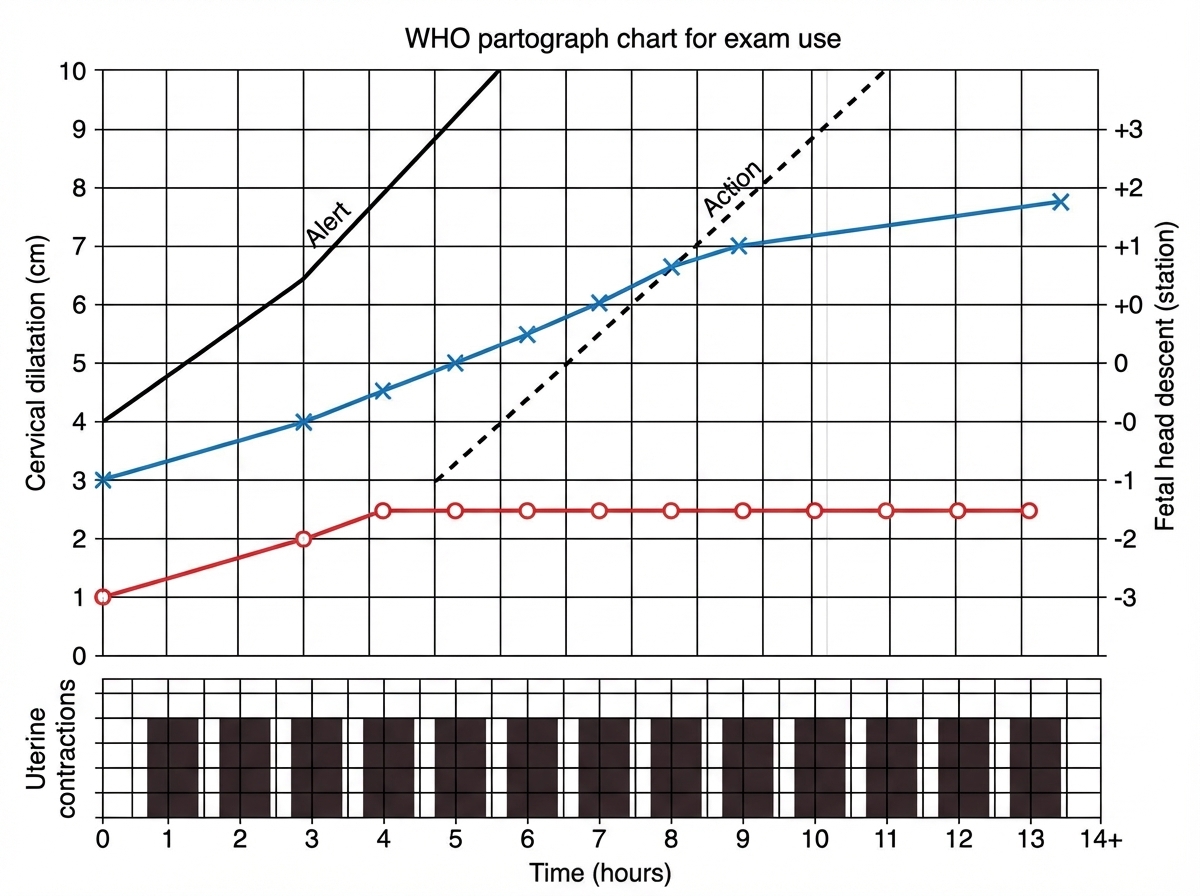
Interpret the given partogram:

Explanation: **Explanation:** The **Occipitoposterior (OP) position** is the most common malposition encountered during labor. The correct management is **"Wait and Watch"** because approximately **90% of OP positions rotate spontaneously** to the occipito-anterior (OA) position during the first and second stages of labor, leading to a normal vaginal delivery. **Why the other options are incorrect:** * **External Cephalic Version (ECV):** This is indicated for converting a breech or transverse lie to a cephalic presentation before labor (usually at 36–37 weeks). It is not used for correcting malpositions like OP. * **Internal Podalic Version:** This is a high-risk procedure used almost exclusively for the delivery of a second twin in a non-vertex presentation. It is contraindicated in singleton cephalic presentations. * **Caesarean Section:** While OP positions are associated with a higher risk of C-section due to "persistent OP" or deep transverse arrest, it is not the primary management. Surgery is reserved for cases where there is failure to progress or fetal distress. **NEET-PG High-Yield Pearls:** 1. **Mechanism:** Rotation occurs due to the well-flexed head meeting the resistance of the pelvic floor (gutter-shaped levator ani). 2. **Persistent OP:** If the head fails to rotate, it results in a "Face-to-Pubes" delivery. 3. **Clinical Sign:** On per-vaginal examination, the anterior fontanelle (diamond-shaped) is felt anteriorly, and the sagittal suture is in the oblique diameter. 4. **Associated Risks:** Prolonged second stage, increased need for oxytocin, and higher incidence of perineal tears.
Explanation: **Explanation:** Fetal distress (non-reassuring fetal status) occurs when the fetus is subjected to hypoxia and acidosis. The goal of monitoring is to identify these changes early to prevent permanent damage or stillbirth. **Why Option D is the correct answer:** Fetal scalp blood sampling is the gold standard for assessing fetal hypoxia. A **pH of 7.3 is considered normal** and reassuring. In the context of fetal distress, the pH typically drops. The clinical thresholds are: * **Normal:** >7.25 * **Pre-acidotic (Borderline):** 7.20 – 7.25 * **Acidosis (Distress):** <7.20 **Analysis of other options:** * **A. Meconium staining:** While not always indicative of distress in post-term pregnancies, the passage of meconium in utero often results from vagal stimulation due to umbilical cord compression or hypoxia, making it a classic sign of distress. * **B. Heart rate < 100 bpm:** Fetal bradycardia (baseline <110 bpm) is a significant indicator of distress. A rate below 100 bpm, especially if persistent or associated with late decelerations, suggests severe fetal compromise. * **C. Involuntary muscle movements:** This refers to "fetal gasping" or excessive, tumultuous fetal movements followed by a decrease in activity, which occurs as a response to acute hypoxia. **NEET-PG High-Yield Pearls:** 1. **Normal Fetal Heart Rate:** 110–160 bpm. 2. **Early Decelerations:** Due to head compression (Physiological/Benign). 3. **Late Decelerations:** Due to uteroplacental insufficiency (Pathological). 4. **Variable Decelerations:** Due to cord compression (Most common type). 5. **Amniotic Fluid Index (AFI):** Normal is 5–24 cm; <5 cm indicates oligohydramnios, often associated with chronic fetal distress.
Explanation: **Explanation:** In a face presentation, the position is determined by the location of the **mentum (chin)**. In a **Mento-Posterior (MP)** position, the fetal head is already in a state of maximum extension. For a vaginal delivery to occur, the head must undergo further extension to sweep over the perineum; however, since the head is already fully extended, no further extension is anatomically possible. Furthermore, the short neck of the fetus cannot span the length of the maternal sacrum (approx. 12 cm), causing the fetal thorax to enter the pelvis simultaneously with the head, leading to **persistent wedge-shaped engagement** and obstructed labor. **Why the other options are incorrect:** * **Vaginal Delivery (A):** Spontaneous vaginal delivery is physically impossible in a persistent mento-posterior position because the head cannot flex or extend further to navigate the pelvic curves. * **Forceps Delivery (B):** Application of forceps is strictly contraindicated in MP positions as it can lead to severe maternal trauma and fetal cervical spine injury without achieving descent. * **Manual Rotation (C):** Manual or forceps rotation from MP to Mento-Anterior (MA) is no longer recommended in modern obstetrics due to high risks of uterine rupture and fetal cord prolapse. **Clinical Pearls for NEET-PG:** * **Mento-Anterior (MA):** Can be delivered vaginally (the chin can pivot under the symphysis pubis). * **Mento-Posterior (MP):** "The chin is back, the baby is stuck." Immediate **Cesarean Section** is the management of choice. * **Internal Podalic Version:** This is contraindicated in face presentations. * **Commonest cause:** Prematurity or anencephaly (due to lack of vertex formation).
Explanation: **Explanation:** **1. Why Transverse Lie is Correct:** Cord prolapse occurs when the umbilical cord descends below the presenting part after the rupture of membranes. The primary risk factor is a **poor fit** between the presenting part and the lower uterine segment/pelvic inlet. In a **transverse lie**, the pelvic inlet is completely unoccupied by a fetal pole, leaving a large space for the cord to prolapse. Statistically, transverse lie carries the highest relative risk (up to 20%) compared to other malpresentations. **2. Analysis of Incorrect Options:** * **B. Breech presentation:** While breech is a common cause of cord prolapse (especially footling breech due to irregular filling of the cervix), the incidence is lower than in transverse lie. * **C. Contracted pelvis:** This prevents the head from engaging (cephalopelvic disproportion), creating space for the cord. However, it is a mechanical factor rather than a primary malpresentation. * **D. Prematurity:** Premature babies are smaller and often associated with malpresentation and polyhydramnios, increasing risk. However, the anatomical "gap" created by a transverse lie remains the most potent risk factor. **3. Clinical Pearls for NEET-PG:** * **Most common risk factor (Overall):** Rupture of membranes (especially artificial rupture/ARM) when the head is high. * **Most common presentation associated:** Cephalic (simply because it is the most common presentation), but the **highest risk/most common association per case** is Transverse Lie. * **Management:** Immediate action is to displace the presenting part upwards (manual elevation or Trendelenburg/Knee-chest position) and perform an emergency Cesarean section. * **Diagnosis:** Sudden fetal bradycardia or variable decelerations following the rupture of membranes.
Explanation: **Explanation:** The third stage of labor begins after the delivery of the fetus and ends with the expulsion of the placenta. The diagnosis in this case is **Retained Placenta**, which is clinically defined as the failure of the placenta to be expelled within **30 minutes** of the birth of the baby (with active management). **Why Option B is correct:** Once 30 minutes have elapsed, the risk of postpartum hemorrhage (PPH) increases significantly. The definitive management for a retained placenta is **Manual Removal of Placenta (MROP)**. This procedure is performed under general anesthesia or deep sedation. The clinician follows the umbilical cord to locate the placenta and uses a "sawing" motion with the edge of the hand to shear the placenta from the uterine wall. **Why other options are incorrect:** * **A. Controlled Cord Traction (CCT):** This is a component of Active Management of the Third Stage of Labor (AMTSL). If the placenta is not delivered within 30 minutes despite CCT, further traction carries the risk of uterine inversion or cord avulsion. * **C. Hysterotomy:** This is a major surgical procedure and is only indicated in cases of *Placenta Accreta Spectrum* where MROP fails or causes massive hemorrhage. * **D. Tocolytics:** These drugs relax the uterus. They are contraindicated here as a contracted uterus is necessary to prevent bleeding. Conversely, *uterotonics* (like Oxytocin) are used after MROP to ensure uterine contraction. **High-Yield Clinical Pearls for NEET-PG:** * **Time limit:** 30 minutes (Active management); 60 minutes (Physiological management). * **Most common cause:** Uterine atony or a constricted "hourglass" contraction ring. * **Prophylaxis:** AMTSL (Oxytocin 10 IU IM) reduces the incidence of retained placenta. * **Post-procedure:** Always check for placental completeness and administer antibiotics to prevent endomyometritis.
Explanation: **Explanation:** Placenta previa occurs when the placenta implants in the lower uterine segment, partially or completely covering the internal os. **Why Option C is the correct answer (The False Statement):** Contrary to common belief, **premature labor is actually infrequent** in placenta previa. While "preterm delivery" is very common, it is usually **iatrogenic** (physician-induced) due to heavy bleeding necessitating emergency delivery, rather than spontaneous onset of labor. The lower segment is passive, and the presence of the placenta there does not typically trigger the biochemical cascade of labor. **Analysis of Incorrect Options (True Statements):** * **Option A:** Postpartum hemorrhage (PPH) is a major risk. The lower uterine segment is less muscular and lacks the "living ligatures" (interlocking muscle fibers) found in the fundus. Consequently, it cannot contract effectively to compress vessels after placental separation. * **Option B:** While the classic presentation is painless bleeding in the third trimester (warning hemorrhage), early "threatened abortion" or first-trimester spotting is not uncommon due to the low implantation. * **Option C:** There is a strong correlation between a scarred uterus (previous LSCS or myomectomy) and placenta previa. The risk increases linearly with the number of previous cesarean sections. **NEET-PG High-Yield Pearls:** * **Classic Presentation:** Painless, causative, recurrent, bright red vaginal bleeding. * **Stallworthy’s Sign:** Posterior placenta previa prevents the head from engaging, causing the fetal heart rate to drop when the head is pushed into the pelvis (due to cord compression). * **Investigation of Choice:** Transvaginal Ultrasound (TVS) is the gold standard (safer and more accurate than transabdominal). * **Contraindication:** **Never** perform a per-vaginal (PV) examination in a suspected case of placenta previa, as it can provoke torrential hemorrhage.
Explanation: **Explanation:** The application of forceps requires specific prerequisites to ensure maternal and fetal safety. The correct answer is **Option C** because forceps application is contraindicated when the presenting part is at **zero station** (the level of the ischial spines). **1. Why Option C is the correct answer:** According to the ACOG classification, forceps application requires the fetal head to be at least at **+2 station** or lower. A station of "zero" is considered a **High Forceps** procedure (if attempted), which is strictly contraindicated in modern obstetrics due to the high risk of maternal trauma (perineal tears, uterine rupture) and fetal injury (intracranial hemorrhage). Forceps should only be applied when the head is engaged and low in the pelvis. **2. Analysis of other options:** * **Option A:** Forceps can be applied when the head is in the **Low Forceps** position (station ≥ +2) even if rotation is <45°. A 15-degree deviation is easily corrected during the traction process. * **Option B:** In a **Face presentation**, forceps are indicated only if the position is **Mento-Anterior**. (Note: Mento-posterior is a contraindication as the head cannot flex to deliver). * **Option C:** The presence of **Caput Succedaneum** is not a contraindication. However, the clinician must be careful to identify the actual bony station of the skull, as a large caput can give a false impression that the head is lower than it actually is. **High-Yield Clinical Pearls for NEET-PG:** * **Prerequisites for Forceps (Mnemonic: FORCEPS):** **F**etus alive, **O**s fully dilated, **R**uptured membranes, **C**ephalopelvic disproportion absent, **E**ngaged head, **P**elvis adequate, **S**ubstantial anesthesia/Empty bladder. * **Station Requirement:** Outlet forceps (Scalp visible at introitus); Low forceps (Station ≥ +2); Mid-forceps (Station 0 to +2, but engaged). * **Most common indication:** Fetal distress or prolonged second stage of labor.
Explanation: The **mid-pelvis** (plane of least pelvic dimensions) is the most critical area of the pelvic canal because it is where the fetal head typically undergoes internal rotation. ### **Explanation of the Correct Answer** The **transverse diameter** of the mid-pelvis is also known as the **Interspinous Diameter**. It is the distance between the two ischial spines. In a standard female pelvis, this measures **10.5 cm**. It is clinically significant because it is the narrowest diameter of the entire pelvic canal through which the fetus must pass. ### **Analysis of Incorrect Options** * **B. 11.5 cm:** This is the approximate **Anteroposterior (AP) diameter** of the mid-pelvis (measured from the lower border of the symphysis pubis to the junction of the 4th and 5th sacral vertebrae). * **C. 12.5 cm:** This corresponds to the **Transverse diameter of the Pelvic Inlet**, which is the widest diameter of the inlet. * **D. 9.5 cm:** This is the **Obstetric Conjugate** (the narrowest AP diameter of the inlet) or the **Bispinous diameter** in a severely contracted pelvis. ### **High-Yield Clinical Pearls for NEET-PG** * **Boundaries of Mid-pelvis:** Anteriorly by the lower border of the symphysis pubis, laterally by the ischial spines, and posteriorly by the sacrum (S4–S5). * **Clinical Significance:** If the interspinous diameter is **<10 cm**, it suggests mid-pelvic contraction, which may lead to transverse arrest of the fetal head. * **Ischial Spines:** These serve as the landmark for "Zero Station" in assessing fetal descent. * **Shape:** The mid-pelvis is roughly circular, but the interspinous diameter is its most restrictive dimension.
Explanation: ### Explanation The correct diagnosis is **Threatened Abortion**. This clinical scenario is defined by vaginal bleeding occurring before the 20th week of gestation where the pregnancy continues. **1. Why Threatened Abortion is correct:** The hallmark of threatened abortion is **vaginal bleeding** with a **closed cervical os** and a uterine size that **matches the period of amenorrhea**. The fetus is typically alive, and the process is potentially reversible. **2. Why the other options are incorrect:** * **Septic Abortion:** This involves an abortion (of any type) complicated by pelvic infection. It presents with fever, malodorous vaginal discharge, and uterine tenderness, which are absent here. * **Complete Abortion:** In this case, the entire products of conception have been expelled. While the os is closed, the **uterine size would be smaller** than the period of amenorrhea, and bleeding would have significantly subsided. * **Inevitable Abortion:** The key differentiator here is the cervix. In inevitable abortion, the **cervical os is dilated**, and the patient usually experiences rupture of membranes or more severe pain/bleeding, making the progression to miscarriage certain. **3. NEET-PG High-Yield Pearls:** * **Management:** Treatment for threatened abortion is primarily **bed rest** (though evidence is limited) and **progesterone supplementation** (to support the corpus luteum). * **Ultrasonography:** It is the investigation of choice to confirm fetal viability. * **Prognosis:** Approximately 50% of threatened abortions progress to inevitable abortion. * **Differential Diagnosis:** Always rule out Ectopic Pregnancy (presents with pain and adnexal mass) and Cervical Polyps.
Explanation: ***Cephalopelvic disproportion*** - The partogram shows **active phase arrest** with the cervical dilatation curve crossing to the right of the **action line**, indicating obstructed labor despite adequate uterine contractions. - This pattern is characteristic of **mechanical obstruction** where the fetal head cannot pass through the maternal pelvis due to size discrepancy. *Inadequate uterine contraction* - Would show **slow cervical dilatation** but the contraction pattern would be weak or infrequent on the partogram. - The cervical dilatation curve would progress slowly but parallel to the **alert line** rather than crossing the action line. *Prolonged latent phase* - Occurs when the **latent phase** (0-4 cm dilatation) takes longer than 20 hours in primigravida or 14 hours in multigravida. - The partogram would show delayed onset of active phase rather than arrest during the **active phase**. *Normalogram* - Would show cervical dilatation progressing at **1 cm/hour** in the active phase, staying to the left of the alert line. - **Fetal heart rate**, **liquor color**, and **descent** would all remain within normal parameters throughout labor.
Physiology of Labor
Practice Questions
Stages of Labor and Normal Progression
Practice Questions
Fetal Monitoring Techniques
Practice Questions
Pain Management in Labor
Practice Questions
Induction and Augmentation of Labor
Practice Questions
Operative Delivery (Forceps and Vacuum)
Practice Questions
Cesarean Section: Indications and Techniques
Practice Questions
Dystocia and Abnormal Labor Patterns
Practice Questions
Obstetric Emergencies
Practice Questions
Postpartum Hemorrhage Management
Practice Questions
Get full access to all questions, explanations, and performance tracking.
Start For Free