The procedure being performed is called:

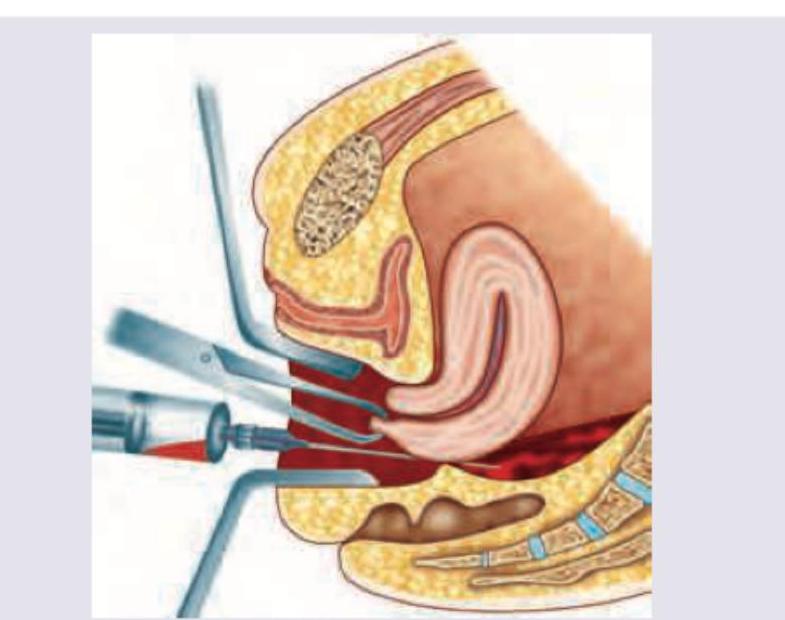
The image shows: (Recent Neet Pattern 2016-17)

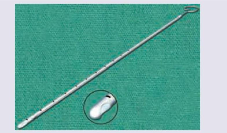
Identify the gynecological instrument shown below. (Recent Neet Pattern 2016-17)

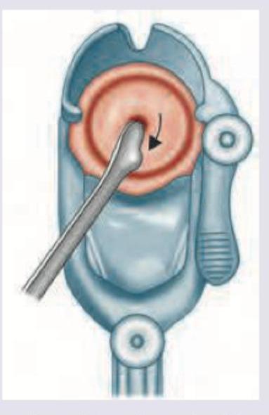
The procedure shown below is called:

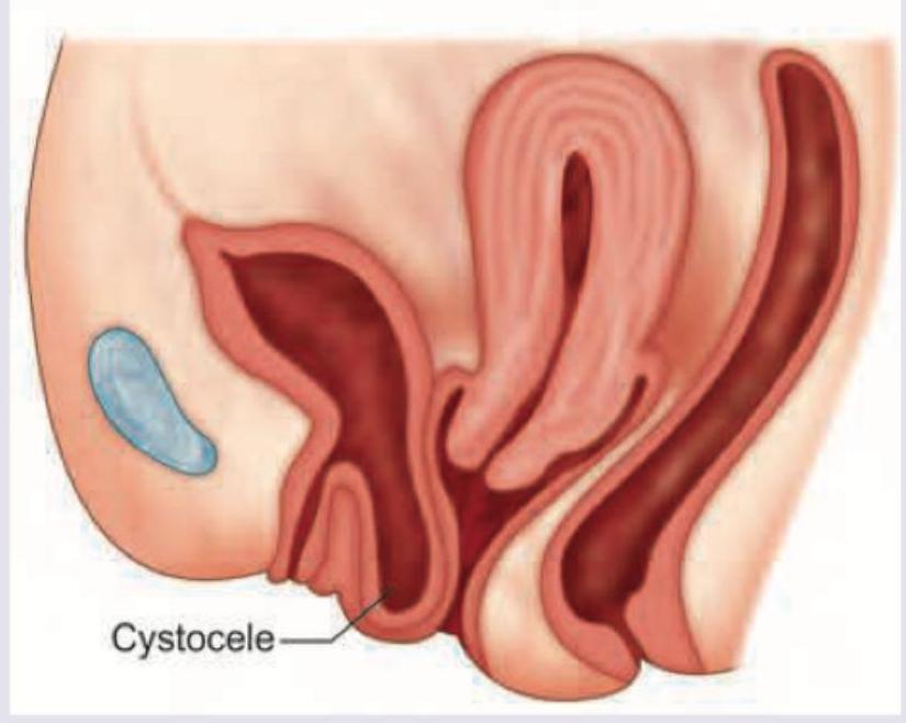
A patient is having following clinical presentation. What is the most likely diagnosis of this condition? (Recent Neet Pattern 2016-17)

All are true about the procedure being performed except: (Recent Neet Pattern 2016-17)

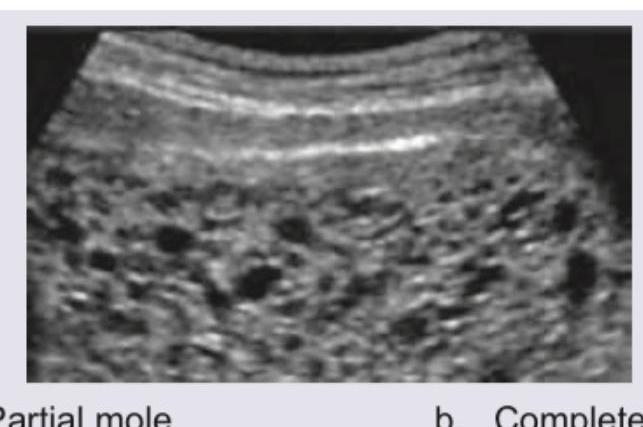
A 25-year-old presents with amenorrhea of 8 weeks. What does the USG show?

What is the best treatment for the condition shown below?

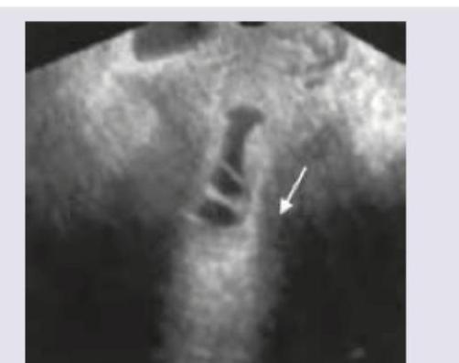
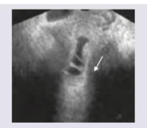
Identify the type of sonography shown:

Transvaginal saline infusion sonographic evaluation shows:

Explanation: **Culdocentesis** - The image depicts a needle being inserted into the **posterior cul-de-sac** (pouch of Douglas) through the posterior vaginal fornix to aspirate fluid, which is the definition of **culdocentesis**. - This procedure is commonly used to detect the presence of **blood** or **pus** in the peritoneal cavity, particularly in cases of suspected ectopic pregnancy or pelvic infection. *Cervicopexy* - **Cervicopexy** is a surgical procedure to fix the cervix in a higher position, typically to prevent or treat uterine prolapse. - It involves suspending the cervix and does not involve fluid aspiration through the vaginal wall. *Cervical cytology* - **Cervical cytology**, also known as a Pap smear, involves collecting cells from the surface of the cervix to screen for **precancerous** or **cancerous changes**. - This procedure uses a brush or spatula to collect cells, not a needle for fluid aspiration from the cul-de-sac. *Colposuspension* - **Colposuspension** is a surgical procedure to treat **stress urinary incontinence** by lifting and supporting the bladder neck and urethra. - This procedure involves a surgical approach through the abdomen, not a transvaginal needle aspiration from the posterior cul-de-sac.
Explanation: ***Uterine sound*** - This instrument is a **uterine sound**, used to measure the **depth of the uterine cavity** before procedures like IUD insertion or D&C. - It is characterized by its long, thin, malleable shaft with markings and usually a handle or ring at one end. *Endometrial curette* - An endometrial curette is typically a **spoon-shaped instrument** used for scraping the uterine lining to obtain tissue samples. - It does not have the uniform, slender, and marked appearance of the instrument shown. *Rocket endocervical brush* - An endocervical brush is designed to collect **cervical cells** and consists of bristles at the tip, which is not depicted here. - This instrument is too rigid and lacks the brush-like tip for cell collection. *Endometrial sampler* - An endometrial sampler, like a Pipelle, is a thin, flexible device with a plunger used to **aspirate endometrial tissue**. - While used for obtaining uterine samples, its structure, particularly the presence of a plunger and suction mechanism, differs from the simple measuring device shown.
Explanation: **Rocket endocervical brush** - The image distinctly shows a **rocket-shaped head** with multiple lamellae, which is characteristic of the Rocket endocervical brush. - This instrument is designed for **simultaneous collection of both ectocervical and endocervical cells** for Pap testing. *Ayre spatula* - An Ayre spatula is typically made of **wood or plastic**, has a flat, broad blade with a notch at one end. - Its design is primarily for **ectocervical scraping**, not the extensive endocervical sampling offered by the pictured brush. *Cervical brush* - While this is a type of cervical brush, the term "cervical brush" is a **general category**. The specific design depicted (rocket-shaped) points to a more precise identification. - A standard cervical brush often has a **single set of bristles** arranged circularly, unlike the multi-lamellar, winged design shown. *Wooden spatula* - A wooden spatula, like the Ayre spatula, is primarily used for **ectocervical cell collection** and has a flat, paddle-like structure. - It lacks the **bristled or lamellar design** that characterizes the instrument shown for simultaneous comprehensive sampling.
Explanation: ***Cone biopsy*** - The image clearly depicts a **cone-shaped section** being removed from the **cervix**, which is characteristic of a cone biopsy (also known as conization). - This procedure is performed to diagnose or treat abnormal cervical cells, typically identified during a Pap test. *Colpocleisis* - This procedure involves **surgically closing the vaginal canal**, often performed for severe pelvic organ prolapse in women who no longer desire vaginal intercourse. - The image shows a cervical excision, not a closure of the vagina. *Cervicopexy* - **Cervicopexy** is a surgical procedure to lift and suspend the cervix, typically to correct **pelvic organ prolapse**. - The illustration shows a fragment of the cervix being removed, not repositioned or suspended. *Colposuspension* - **Colposuspension** is a surgical procedure to support the **bladder neck** and **urethra** to treat **stress urinary incontinence**. - This involves attaching the vaginal wall near the urethra to ligaments in the pelvis, which is different from the cervical tissue removal shown.
Explanation: ***Gartner cyst*** - The image shows a fluid-filled lesion in the **anterolateral vaginal wall**, consistent with a Gartner's cyst, which is a remnant of the **Wolffian duct**. - This cyst is typically located along the course of the Gartner's duct and can be palpated or visualized during pelvic examination. *Rectocele* - A rectocele involves the **posterior vaginal wall bulging into the vaginal lumen** due to rectovaginal fascia weakness, where the rectum protrudes. - The image clearly depicts an anterior vaginal wall lesion, not a posterior one. *Nabothian cyst* - **Nabothian cysts** are retention cysts located on the **cervix**, resulting from blocked cervical glands. - The lesion in the image is located in the vagina, not on the cervix. *Submucous fibroids* - **Submucous fibroids** are **uterine leiomyomas** that protrude into the endometrial cavity, originating from the myometrium. - The image shows a vaginal lesion, not a uterine pathology.
Explanation: ***Procedure to be done after bimanual examination*** - This statement is **FALSE** and is the correct answer to this "except" question - The standard sequence for pelvic examination is: **speculum exam → Pap smear collection → bimanual examination** - The Pap smear must be performed **BEFORE** the bimanual examination because the lubricant used during bimanual examination would contaminate the cervical sample and interfere with cytological interpretation - Performing the procedure after bimanual examination would compromise the quality of the cervical cell collection *Cusco speculum introduced without lubrication* - This statement is **TRUE** in the context of Pap smear collection - When performing a Pap test, the Cusco (Cusco's) speculum should be introduced using **warm water only** as a lubricant, avoiding gel-based or petroleum-based lubricants - Commercial lubricating gels can interfere with the cytological examination and obscure cellular details - While technically using "warm water," this is standard practice for Pap smear procedures *Ayre spatula* - This statement is **TRUE** - The Ayre spatula is the classic wooden spatula designed specifically for collecting ectocervical cells during Pap smear procedures - The spatula has a curved end that fits the contour of the cervix, with the longer projection inserted into the external os - Modern variations include combination devices like cytobrush for endocervical sampling or broom-like devices for liquid-based cytology *Scraping material fixed over slide using 95% ether and alcohol* - This statement is **TRUE** for conventional Pap smear technique, though somewhat outdated - Immediate fixation is crucial to prevent air-drying artifacts - **95% ethyl alcohol** is the standard fixative for conventional Pap smears - While ether-alcohol mixtures were historically used, modern practice typically uses 95% ethanol alone or commercial cytology fixative sprays - Many laboratories now use liquid-based cytology (LBC) where cells are rinsed into a preservative solution, eliminating immediate slide fixation
Explanation: ***Partial mole*** - The ultrasound image shows a **thickened placenta** with **multiple cystic spaces** (vesicles), often described as a "grape-like cluster" or "Swiss cheese" appearance, which is characteristic of a partial hydatidiform mole. - In a partial mole, there is often **fetal tissue present**, which may be seen as a small, abnormal fetus or amniotic sac, although sometimes it can be difficult to identify in early stages or if very rudimentary. *Complete mole* - A **complete mole** would typically show a **uterus filled with hydropic villi** and no evidence of fetal parts or an amniotic sac. - The "snowstorm" appearance due to diffuse hydropic villi is more characteristic of a complete mole, and while there are vesicles here, the presence of what appears to be some organized placental tissue with vesicles within it is more typical of a partial mole. *PCOS* - **Polycystic Ovary Syndrome (PCOS)** is characterized by **multiple small follicles (cysts)** arranged peripherally in the ovaries, and it would not present with this kind of uterine contents, which is clearly a product of conception. - PCOS is a hormonal disorder leading to chronic anovulation and hyperandrogenism, and its ultrasound findings are distinct from placental abnormalities. *Endometrioma* - An **endometrioma** is a type of **ovarian cyst** filled with old blood, presenting as a **homogenous, hypoechoic mass** with internal echoes, often described as a "ground glass" appearance. - This image clearly depicts contents within the uterine cavity related to a pregnancy, not an ovarian cyst.
Explanation: ***Hysteroscopic adhesiolysis*** - The image shows an **intrauterine synechia**, also known as an **Asherman syndrome** or uterine adhesion, which manifests as fibrous bands within the uterine cavity (indicated by the arrow). - **Hysteroscopic adhesiolysis** is the gold standard for treating intrauterine adhesions, involving the surgical removal of these adhesions under direct visualization to restore the uterine cavity's normal anatomy. *OCP* - **Oral contraceptive pills (OCPs)** are primarily used for contraception, managing menstrual irregularities, or hormonal therapy. - They do not address or resolve **intrauterine adhesions** causing conditions like Asherman syndrome. *Transvaginal hysterectomy* - A **transvaginal hysterectomy** involves the surgical removal of the entire uterus, which is an aggressive and irreversible procedure. - This is not an appropriate treatment for intrauterine adhesions, especially in patients who may desire future fertility. *Intrauterine hyaluronidase injection* - While hyaluronidase is an enzyme that breaks down hyaluronic acid and has been explored in various applications, its efficacy as a primary treatment for established **intrauterine adhesions** is not well-supported in clinical practice. - The **mechanical removal** of fibrous adhesions is typically required.
Explanation: ***Saline infusion sonography*** - The image shows a **fluid-filled uterine cavity** (hypoechoic space) with a visible **echogenic endometrial lining**, which is characteristic of saline infusion sonography. - This technique involves injecting saline into the uterus to distend the cavity, allowing for better visualization of **endometrial abnormalities** like polyps or fibroids. *Doppler sonography* - **Doppler sonography** is used to assess **blood flow** and would show color overlays or spectral patterns indicating flow direction and velocity, which are absent here. - It's often used to evaluate **vascularity of masses** or blood flow in vessels, not to outline the uterine cavity with fluid. *3D sonography* - **3D sonography** generates **multiplanar views** and volumetric images, creating a three-dimensional reconstruction, which is not depicted in this single 2D image. - While it can be used to evaluate uterine anomalies, this particular image does not show the typical output of a 3D reconstruction. *Hysterosalpingography* - **Hysterosalpingography (HSG)** involves injecting **radiopaque contrast** into the uterus and fallopian tubes, followed by X-ray imaging. - It would appear as a series of X-ray images, with contrast visible in the tubes and spilling into the peritoneum, not an ultrasound image of the uterine cavity.
Explanation: ***Asherman's syndrome*** - The image shows **intracavitary adhesions** within the endometrial cavity, characterized by bridging bands or strands of tissue with an irregular endometrial surface, as indicated by the arrow. - These findings on **saline infusion sonography (SIS)** are highly suggestive of **Asherman's syndrome**, which involves the formation of intrauterine adhesions. *Submucous leiomyoma* - Submucous leiomyomas (submucosal fibroids) are fibroids that project into the endometrial cavity and typically appear as **well-defined, hypoechoic masses** that distort the cavity. - The image does not show a distinct mass but rather **irregular strands** and a distorted cavity indicative of adhesions, not a fibroid. *Cervical stenosis* - Cervical stenosis involves an **abnormal narrowing of the cervical canal**, which would be seen as an obstruction to fluid flow into the uterine cavity during SIS. - While the image shows fluid within the uterine cavity, the primary pathology is within the cavity itself, not merely an obstruction at the cervix. *PCOD* - **Polycystic ovary syndrome (PCOS)** is primarily an ovarian disorder characterized by **multiple small follicles** (cysts) in the ovaries and hormonal imbalances. - PCOS is not a condition diagnosed by evaluating the endometrial cavity for adhesions using saline infusion sonography.
Abnormal Uterine Bleeding
Practice Questions
Endometriosis
Practice Questions
Adenomyosis
Practice Questions
Uterine Fibroids
Practice Questions
Ovarian Cysts
Practice Questions
Pelvic Inflammatory Disease
Practice Questions
Vulvovaginitis
Practice Questions
Pelvic Organ Prolapse
Practice Questions
Vulvar Disorders
Practice Questions
Benign Breast Diseases
Practice Questions
Get full access to all questions, explanations, and performance tracking.
Start For Free