Which of the following is incorrect about the instrument shown below? (Recent Neet Pattern 2016-17)

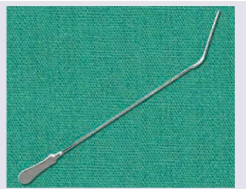
What is the instrument shown below used for?

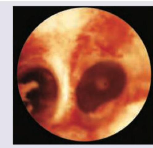
The following hysteroscopy view shows presence of: (Recent Neet Pattern 2016-17)

A 23-year-old woman presents to your clinic with complaints of vulvar pain. The patient's history is significant for a new sexual partner and a recent history of flu-like symptoms and vaginal burning. On physical examination, extremely painful shallow ulcers with red borders are appreciated on the vulva, vagina, and perineal region. Which of the following is the most appropriate course of treatment for this patient?
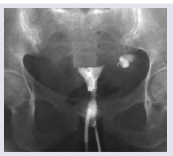
HSG image given below shows:

A 19-year-old woman presents to the emergency department complaining of lesions on her vulva. They have been present for several months but are now beginning to interfere with intercourse. Which viral subtypes are responsible for most cases of this disease? (Recent Neet Pattern 2016-17)
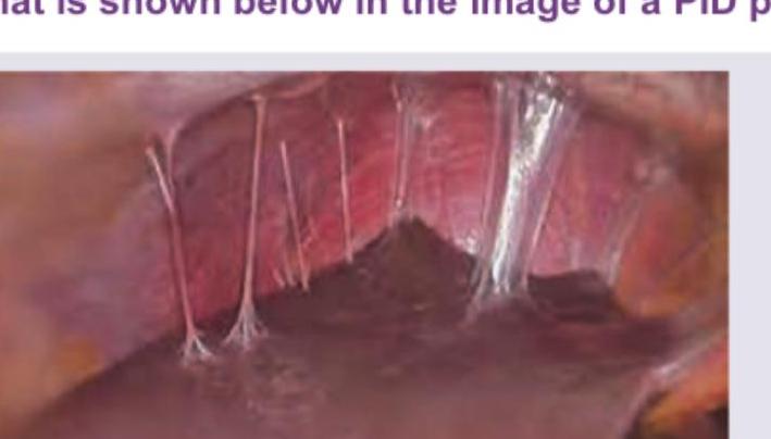
What is shown below in the image of a PID patient?

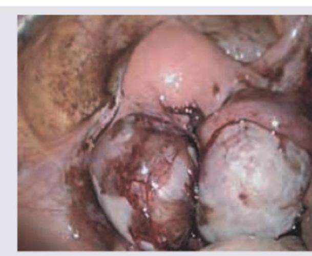
A 28 -year-old female patient presented with lower abdominal pain along with dysmenorrhea. The following finding was seen on laparoscopic examination. What is the likely diagnosis?

A 21-year-old girl is having vaginal discharge one week after unprotected sexual intercourse. The smear of vaginal discharge is shown below. All are diagnostic criteria for the condition shown except:

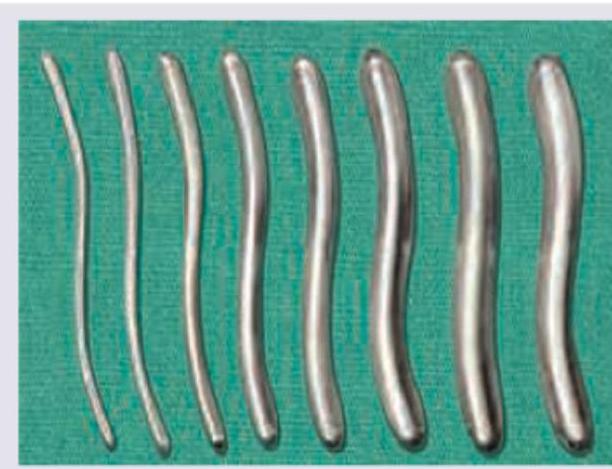
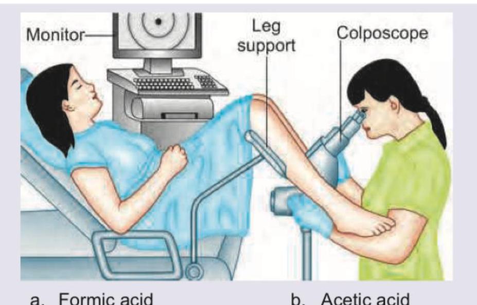
Which of the following is used in the gynecological procedure being performed in the image?

Explanation: ***Contraindicated in postpartum state*** - The instrument shown is a **uterine sound**, used to measure the **depth of the uterine cavity**. It is **not contraindicated in the postpartum state**; in fact, it can be used cautiously to check for retained products of conception or assess the uterine cavity, keeping in mind the uterus is softer and more prone to perforation. - This statement is **INCORRECT**, making it the right answer to this question. *Used to determine uterine size* - A **uterine sound** is primarily used to measure the **length of the uterine cavity from the external os to the fundus**, which helps determine uterine size. - This measurement is crucial for procedures like **IUD insertion**, endometrial biopsy, and hysteroscopy. - This is a **CORRECT** statement about the uterine sound. *Passed till it meets resistance at the fundus* - The uterine sound is passed through the **cervical canal** until it gently touches the **fundus** (top) of the uterus. - The measurement is read from the marking on the instrument corresponding to the external cervical os, typically ranging from **6-8 cm** in normal uteri. - This is a **CORRECT** statement about the technique. *Malleable sound* - The image depicts a **uterine sound**, which is typically made of **malleable metal** (usually stainless steel). - Its malleability allows it to be bent or shaped to match the **curvature of the uterus** (anteversion/retroversion), reducing the risk of perforation and facilitating accurate measurement. - This is a **CORRECT** statement about the instrument.
Explanation: ***Dilating the ostium uteri*** - The image depicts a set of **Hegar dilators**, which are instruments used to **dilate the cervical os (ostium uteri)**. - They come in graduated sizes and are inserted sequentially to gradually widen the cervical canal for procedures like D&C, IUD insertion, or hysteroscopy. *Dilate urethral strictures* - While dilators are used for urethral strictures, the instruments shown are specifically designed with a curvature and length optimized for the **cervical canal**, not the male or female urethra. - **Urethral stricture dilators** generally have a different shape, often longer and straighter or with a specific angle for the male urethra. *Measure uterus size in pregnancy* - Measuring uterus size in pregnancy is typically done by **fundal height measurement** with a tape measure or **ultrasound**, not with these solid dilators. - Hegar dilators are for dilating, not measuring length or size of the uterus during pregnancy. *Measure thickness of uterine wall* - The thickness of the uterine wall is measured using **imaging techniques** like **ultrasound** or MRI. - These dilators are solid instruments designed for mechanical dilation and cannot measure tissue thickness.
Explanation: ***Septate uterus*** - The image clearly shows a **distinct septum** dividing the **uterine cavity** into two hemicavities, a characteristic feature of a septate uterus. - This anatomical anomaly is a common cause of **recurrent pregnancy loss** and can be resected hysteroscopically. *Normal uterine cavity* - A normal uterine cavity would appear as a **single, smooth, triangular space** without any dividing structures. - The presence of a **central wall** clearly indicates an abnormality, ruling out a normal cavity. *Asherman syndrome* - Asherman syndrome is characterized by the presence of **intrauterine adhesions** or **synechiae**, which are fibrous bands often appearing as thin, web-like structures or dense scar tissue. - While adhesions can divide the cavity, they typically appear as **irregular, often avascular, fibrous strands** rather than a well-formed, fleshy septum as seen here. *Uterine perforation* - Uterine perforation would typically present as a **hole or tear in the uterine wall**, which might lead to visualization of extrauterine structures or free fluid in the peritoneal cavity. - The image shows an **internal anatomical division** of the uterus, not a breach of its outer wall.
Explanation: ***Acyclovir*** - The patient's symptoms, including **vulvar pain**, **flu-like symptoms**, **vaginal burning**, and **extremely painful shallow ulcers with red borders** are highly suggestive of a **primary herpes simplex virus (HSV) infection**. - **Acyclovir**, a guanosine analog, is an antiviral medication that specifically inhibits viral DNA synthesis and is the treatment of choice for HSV infections. *Penicillin* - **Penicillin** is an antibiotic primarily used to treat bacterial infections, particularly those caused by gram-positive bacteria like *Streptococcus* and *Staphylococcus*, and certain sexually transmitted infections such as **syphilis**. - The clinical presentation with **vesicular/ulcerative lesions** and **flu-like prodrome** is not consistent with bacterial infections responsive to penicillin. *Azithromycin* - **Azithromycin** is a macrolide antibiotic commonly used to treat bacterial infections such as **chlamydia**, **gonorrhea**, and certain respiratory tract infections. - The described lesions and systemic symptoms do not fit the typical presentation of bacterial sexually transmitted infections treated with azithromycin. *Doxycycline* - **Doxycycline** is a tetracycline antibiotic effective against a broad range of bacterial infections, including **chlamydia**, **lyme disease**, and **rickettsial infections**. - It is not indicated for **viral infections** like HSV, which is characterized by painful vesicular or ulcerative lesions.
Explanation: ***Asherman syndrome*** - The HSG image shows multiple **filling defects** and an **irregular, partially obliterated endometrial cavity**, which are characteristic findings of Asherman syndrome. - This condition is caused by **intrauterine adhesions** (synechiae) resulting from trauma to the endometrial lining, typically due to procedures like D&C. *Endometrial polyp* - An endometrial polyp would typically appear as a **single, well-defined filling defect** within the endometrial cavity. - The image demonstrates **multiple, irregular adhesions** rather than a solitary, smooth-bordered mass. *Genital TB* - Genital tuberculosis can cause a variety of HSG findings, including **calcifications**, **beading of the fallopian tubes**, and a **"T-shaped" or constricted endometrial cavity**. - While it can cause intrauterine adhesions, the image's appearance of widespread, irregular filling defects with partial obliteration is more classic for Asherman syndrome, specifically if there is no other evidence of TB. *Subserosal fibroids* - Subserosal fibroids are located on the **outer surface of the uterus** and typically do not distort the endometrial cavity or appear as filling defects within it on an HSG. - On HSG, they may cause **extrinsic compression** or displacement of the uterus but would not appear as intraluminal filling defects or adhesions.
Explanation: ***Types 6 and 11*** - Human Papillomavirus (HPV) **types 6 and 11** are responsible for approximately 90% of all cases of **genital warts (condyloma acuminata)**. - These types are considered low-risk HPV as they rarely lead to cancer but can cause significant discomfort and interfere with sexual activity due to their physical presence. *Types 6 and 18* - While **HPV type 6** is a common cause of genital warts, **HPV type 18** is a high-risk type primarily associated with **cervical cancer**, not typically with vulvar warts. - Combining a low-risk and a high-risk type in this manner does not accurately represent the primary cause of condyloma acuminata. *Types 11 and 16* - **HPV type 11** causes genital warts, but **HPV type 16** is a **high-risk HPV** type strongly associated with cervical and other anogenital cancers. - While both can cause lesions, type 16 is not a primary cause of benign genital warts; it's a major cause of malignancies. *Types 11 and 31* - **HPV type 11** is linked to genital warts, but **HPV type 31** is another **high-risk HPV** type associated with cervical cancer, not typically with benign vulvar warts. - Similar to the previous option, this combination incorrectly mixes a benign wart-causing type with a high-risk oncogenic type when addressing the cause of vulvar lesions.
Explanation: ***Fitz-Hugh-Curtis syndrome*** - The image displays **violin string adhesions** between the liver capsule and the parietal peritoneum, which are characteristic of Fitz-Hugh-Curtis syndrome. - This condition is a perihepatitis, often associated with **pelvic inflammatory disease (PID)**, caused by bacterial infection spreading from the pelvic organs to the liver capsule. *Asherman syndrome* - This condition involves the formation of **intrauterine adhesions** causing partial or complete obliteration of the uterine cavity. - Asherman syndrome typically results from trauma to the endometrial lining, such as from dilation and curettage, and is characterized by menstrual abnormalities and infertility, not perihepatic adhesions. *Rokitansky syndrome* - Also known as Mayer-Rokitansky-Küster-Hauser (MRKH) syndrome, this is a congenital disorder characterized by **agenesis of the uterus and vagina**. - It involves developmental abnormalities of the female reproductive tract and does not present with intra-abdominal adhesions. *Keratoderma blennorrhagicum* - This is a mucocutaneous manifestation of **reactive arthritis**, characterized by **hyperkeratotic skin lesions** on the palms and soles. - It is a dermatological condition and does not involve intra-abdominal adhesions or directly relate to PID in the manner of Fitz-Hugh-Curtis syndrome.
Explanation: ***Pelvic Endometriosis*** - The laparoscopic image shows classic findings of **endometriosis**, including **dark, hemorrhagic lesions** (powder-burn or gunshot lesions), adhesions, and potentially **endometriomas** (chocolate cysts) on the ovaries. - The patient's symptoms of **lower abdominal pain** and **dysmenorrhea** are highly characteristic of endometriosis, which involves the presence of endometrial-like tissue outside the uterus. - **Laparoscopy** is the gold standard for diagnosing endometriosis, revealing typical hemorrhagic implants and scarring. *Pelvic Inflammatory Disease* - PID typically presents with **acute pelvic pain**, **fever**, **purulent vaginal discharge**, and **cervical motion tenderness**. - Laparoscopic findings would show **hyperemia**, **purulent exudate**, and **tubo-ovarian abscesses**, not the dark hemorrhagic lesions seen here. - The chronic nature of dysmenorrhea and absence of infectious features make PID unlikely. *Ovarian Dermoid Cyst* - Dermoid cysts are **benign mature cystic teratomas** that appear as smooth, round cystic masses on laparoscopy. - They typically do not cause significant dysmenorrhea unless complicated by torsion or rupture. - The hemorrhagic implants and adhesions seen in the image are not consistent with dermoid cysts. *Adenomyosis* - Adenomyosis involves endometrial tissue within the **myometrium** (uterine muscle wall), causing an **enlarged, boggy uterus**. - Diagnosis is typically made by **ultrasound** or **MRI** showing thickened myometrium with heterogeneous echogenicity. - **Laparoscopy** would not reveal the characteristic dark hemorrhagic lesions seen in endometriosis, as adenomyosis is an intrauterine condition.
Explanation: ***Vaginal pH < 4.5*** - Bacterial vaginosis (BV) is characterized by a **vaginal pH > 4.5**, not less than 4.5. This elevated pH is due to the reduction of normal lactobacilli, which produce lactic acid. - Healthy vaginal pH is typically between 3.8 and 4.5; a pH less than 4.5 would suggest a normal vaginal flora or potentially Candida infection, not BV. *Positive whiff test* - A **positive whiff test**, characterized by a **fishy odor** when 10% potassium hydroxide (KOH) is added to a vaginal discharge sample, is a diagnostic criterion for bacterial vaginosis. - This odor results from the release of volatile amines produced by anaerobic bacteria overgrowing in the vagina. *Clue cells = 20% of cells* - The presence of **clue cells**, which are vaginal epithelial cells covered with bacteria, constitutes a key diagnostic criterion for bacterial vaginosis, especially when they represent **20% or more of the epithelial cells** on a wet mount. - The image provided clearly depicts epithelial cells encrusted with coccobacillary bacteria, characteristic of clue cells. *Presence of more Gardnerella with few lactobacillus* - Bacterial vaginosis is caused by an **overgrowth of anaerobic bacteria**, primarily *Gardnerella vaginalis*, and a **reduction in the protective lactobacilli** that normally dominate the vaginal flora. - This imbalance in the vaginal microbiome is central to the pathophysiology of BV and is a diagnostic feature.
Explanation: ***Acetic acid*** - **Acetic acid** (typically 3-5%) is routinely applied to the cervix during **colposcopy** to highlight abnormal areas. - It causes areas of **dysplasia or cancer** to dehydrate and appear as opaque **acetowhite lesions**, making them visible under magnification. *Formic acid* - **Formic acid** is not typically used in colposcopy or similar gynecological procedures for diagnostic purposes. - It is more commonly found in nature (e.g., ant venom) and has industrial applications. *Trichloroacetic acid* - **Trichloroacetic acid (TCA)** is a corrosive agent used for the treatment of **warts** (including genital warts) and as a chemical peel. - While it can be used in gynecology, its primary role is therapeutic destruction of tissue, not diagnostic visualization during colposcopy. *Picric acid* - **Picric acid** is a highly explosive compound and is generally not used in clinical gynecological procedures. - It has historical uses as a dye and antiseptic, but its toxicity and explosive nature make it unsuitable for modern medical diagnostics or treatment.
Abnormal Uterine Bleeding
Practice Questions
Endometriosis
Practice Questions
Adenomyosis
Practice Questions
Uterine Fibroids
Practice Questions
Ovarian Cysts
Practice Questions
Pelvic Inflammatory Disease
Practice Questions
Vulvovaginitis
Practice Questions
Pelvic Organ Prolapse
Practice Questions
Vulvar Disorders
Practice Questions
Benign Breast Diseases
Practice Questions
Get full access to all questions, explanations, and performance tracking.
Start For Free