Which of the following does NOT predispose to isoimmunization in an Rh-negative female?
What is the mechanism responsible for high rates of spontaneous abortion in a septate uterus?
What is the best investigation to establish the diagnosis of endometriosis?
What is pathognomonic of ectopic pregnancy?
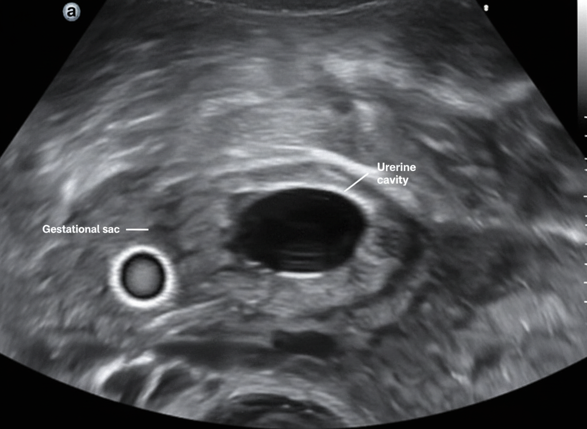
Which of the following conditions is NOT associated with the pathology shown in the ultrasound image?

Which of the following is NOT a complication of hysterectomy?
A 45-year-old postmenopausal woman presents with vaginal bleeding. Transvaginal ultrasound reveals an endometrial thickness of 8.0 mm. What is the next step in management?
Hematocolpos is seen in which of the following conditions?
What is the sensitivity of a conventional Pap smear in detecting cervical cancer?
Curdy vaginal discharge is characteristic of which infection?
Explanation: **Explanation:** Rh isoimmunization occurs when an Rh-negative mother is exposed to Rh-positive fetal red blood cells, leading to the production of maternal antibodies. This process requires a **feto-maternal hemorrhage (FMH)**. **Why Advanced Maternal Age is the Correct Answer:** Advanced maternal age (Option A) is a risk factor for chromosomal abnormalities and certain obstetric complications (like preeclampsia), but it does **not** inherently cause the mixing of maternal and fetal blood. Therefore, it does not predispose a woman to isoimmunization. **Analysis of Incorrect Options:** * **Antepartum Hemorrhage (Option B):** Conditions like abruptio placentae or placenta previa involve the disruption of the placental barrier, which significantly increases the risk of FMH and subsequent sensitization. * **Cesarean Section (Option C):** Any intrauterine surgical intervention, including C-sections and manual removal of the placenta, increases the volume of fetal blood entering the maternal circulation compared to a normal vaginal delivery. * **Postdated Pregnancy (Option D):** In post-term pregnancies, the placenta undergoes physiological aging and degeneration (infarcts, calcifications). This structural weakening makes the placental barrier more "leaky," allowing fetal erythrocytes to cross into the maternal bloodstream even in the absence of trauma. **High-Yield Clinical Pearls for NEET-PG:** * **Minimum blood volume for sensitization:** As little as **0.1 ml** of Rh-positive fetal blood can cause isoimmunization. * **Standard Dose:** 300 mcg of Anti-D IgG can neutralize **15 ml of fetal RBCs** (or 30 ml of whole fetal blood). * **Kleihauer-Betke Test:** Used to quantify the volume of FMH to determine if additional doses of Anti-D are required. * **Routine Immunoprophylaxis:** Administered at **28 weeks** gestation and within **72 hours** of delivery if the neonate is Rh-positive.
Explanation: ### Explanation **Correct Answer: C. Implantation on an avascular septum** The **septate uterus** is the most common Mullerian duct anomaly and is associated with the highest rate of reproductive failure (spontaneous abortions). The primary mechanism is the **poor vascularization** of the septum. The septum is composed of fibroelastic tissue with significantly fewer blood vessels compared to the normal myometrium. If the blastocyst implants directly onto this relatively **avascular septum**, the developing embryo receives inadequate blood supply, leading to early pregnancy loss (typically in the first trimester). **Analysis of Incorrect Options:** * **A. Distorted uterine cavity:** While the cavity is divided, simple distortion is more characteristic of a *bicornuate* or *unicornuate* uterus. In a septate uterus, the external contour is normal; the internal division's primary issue is the quality of the tissue, not just the shape. * **B. Associated cervical abnormality:** Cervical incompetence is more frequently associated with a *bicornuate* or *didelphys* uterus. While it can occur in a septate uterus, it is not the primary cause of the high abortion rate. * **D. Unfavourable endometrial lining:** While the endometrium overlying the septum may be histologically different (asynchronous), the root cause is the underlying lack of blood vessels (avascularity) to support that lining and the subsequent pregnancy. **NEET-PG High-Yield Pearls:** * **Most common Mullerian anomaly:** Septate uterus (also has the worst prognosis for pregnancy). * **Gold Standard for Diagnosis:** Combined Laparoscopy and Hysteroscopy (to differentiate from a bicornuate uterus by checking the external fundal contour). * **Treatment of Choice:** Hysteroscopic Septal Resection (Metzenbaum procedure). * **Distinguishing Feature:** A septate uterus has a **flat or convex** external fundal notch, whereas a bicornuate uterus has a **deep indentation (>1 cm)**.
Explanation: **Explanation:** **Laparoscopy** is considered the **Gold Standard** investigation for the diagnosis of endometriosis. This is because it allows for direct visualization of characteristic lesions (such as "powder-burn" or "mulberry" spots) on the pelvic peritoneum and ovaries. Furthermore, it enables the clinician to perform a biopsy for histopathological confirmation, which is the definitive diagnostic requirement. **Why other options are incorrect:** * **Ultrasound (USG):** While Transvaginal Sonography (TVS) is the first-line imaging modality for detecting **Endometriomas** (chocolate cysts), it has very low sensitivity for identifying superficial peritoneal implants or small adhesions. * **CT Scan:** It has limited soft-tissue resolution in the pelvis and is generally not useful for diagnosing endometriosis, though it may be used to rule out other pathologies. * **X-ray Pelvis:** This is an obsolete investigation for endometriosis as soft tissue implants and cysts are not radio-opaque and cannot be visualized on a plain radiograph. **High-Yield Clinical Pearls for NEET-PG:** * **Definitive Diagnosis:** Histopathology (biopsy) following laparoscopy. * **First-line Investigation:** Transvaginal Ultrasound (TVS). * **MRI:** Useful for diagnosing **Deep Infiltrating Endometriosis (DIE)** involving the rectovaginal septum or bladder. * **Biomarker:** **CA-125** may be elevated in endometriosis, but it is non-specific and used primarily for monitoring recurrence rather than primary diagnosis. * **Classic Triad:** Dysmenorrhea (congestive), Dyspareunia, and Infertility.
Explanation: **Explanation:** **Why Decidual Casts are Correct:** A **decidual cast** is the pathognomonic finding of an ectopic pregnancy. It occurs when the endometrium, which has thickened and become "decidualized" due to rising progesterone levels from the pregnancy, is shed in its entirety as a single, fleshy, triangular piece of tissue. This happens because the ectopic pregnancy cannot sustain the hormonal levels required to maintain the uterine lining. While it mimics a miscarriage, the absence of chorionic villi on histopathological examination (Arias-Stella reaction may be present) confirms that the pregnancy is extrauterine. **Analysis of Incorrect Options:** * **A. Purple hemorrhagic pregnancy in the lower vagina:** This describes a **Choriocarcinoma** or a vaginal metastasis of a gestational trophoblastic neoplasm, not an ectopic pregnancy. * **C. Chadwick sign:** This is a bluish discoloration of the cervix, vagina, and labia caused by increased vascularity. It is a **presumptive sign of pregnancy** (both intrauterine and ectopic) but is not specific or pathognomonic for ectopic pregnancy. * **D. Postcoital hemorrhage:** This is a classic presentation of **Cervical Cancer** or cervical polyps/ectropion, unrelated to the location of a pregnancy. **High-Yield NEET-PG Pearls:** * **Classic Triad:** Amenorrhea, abdominal pain, and vaginal bleeding (present in only 50% of cases). * **Most common site:** Ampulla of the Fallopian tube. * **Arias-Stella Reaction:** Hypersecretory endometrium seen in ectopic pregnancy; it is suggestive but *not* pathognomonic (can occur in IUGR or molar pregnancy). * **Gold Standard Diagnosis:** Transvaginal Ultrasound (TVUS) + Serum β-hCG (Discriminatory zone: 1500–2000 mIU/mL).
Explanation: ***Clomiphene citrate therapy*** - **Clomiphene citrate** is a selective estrogen receptor modulator that induces ovulation by blocking estrogen receptors at the hypothalamic level, but it does **NOT increase the risk of ectopic pregnancy**. - Unlike **gonadotropin therapy** (which does increase ectopic pregnancy risk), clomiphene has a **normal rate of tubal implantation** and does not affect fallopian tube function or motility. *Salpingitis* - **Pelvic inflammatory disease (PID)** and salpingitis cause **tubal scarring** and **adhesions**, leading to impaired tubal motility and increased risk of ectopic pregnancy. - **Chlamydia** and **gonorrhea** infections are major causes, resulting in **tubal damage** that prevents normal embryo transport to the uterus. *Intrauterine contraceptive device (IUCD)* - **IUCD use** increases the risk of ectopic pregnancy by causing **local inflammatory changes** in the endometrium and fallopian tubes. - While IUCDs prevent intrauterine pregnancy more effectively than tubal pregnancy, the **relative risk of ectopic pregnancy** is increased in IUCD users who do conceive. *Plastic procedure on the fallopian tube* - **Tubal reconstructive surgery** (salpingoplasty, salpingostomy) can cause **tubal scarring** and **adhesion formation**, impairing normal tubal function. - **Previous tubal surgery** disrupts the normal **ciliary action** and **muscular contractions** of the fallopian tube, increasing ectopic pregnancy risk.
Explanation: ### Explanation The correct answer is **Bladder hypotonia**. **1. Why Bladder Hypotonia is NOT a complication:** Hysterectomy, particularly radical hysterectomy (Wertheim’s), involves dissection near the pelvic plexus and vesical nerves. Injury to these parasympathetic nerves typically results in **detrusor areflexia or bladder hypertonia/dysfunction** (difficulty emptying), rather than hypotonia. In most standard total abdominal hysterectomies (TAH), the bladder is more prone to direct surgical trauma or postoperative **detrusor instability (urge incontinence)** due to local irritation or loss of support, but hypotonia is not a recognized clinical complication of the procedure. **2. Analysis of Incorrect Options:** * **Ureteric injury (A):** This is a classic and feared complication. The ureter is most vulnerable at three sites: where it crosses the iliac vessels (infundibulopelvic ligament), where it passes under the uterine artery ("water under the bridge"), and at the vesicoureteric junction. * **Vault prolapse (C):** This is a long-term complication caused by the failure to adequately reattach the cardinal and uterosacral ligaments to the vaginal cuff, leading to the descent of the vaginal apex. * **Vaginal cuff dehiscence (D):** This refers to the separation of the vaginal incision. It is more common in laparoscopic hysterectomies compared to vaginal or abdominal routes, often triggered by early resumption of intercourse or infection. **3. NEET-PG High-Yield Pearls:** * **Most common site of ureteric injury:** At the level of the uterine artery (during clamping of the cardinal ligaments). * **Most common visceral injury:** Bladder injury (more common than ureteric injury). * **Prevention:** Intraoperative identification of the ureter and "skeletonization" of vessels are key. * **Vaginal Vault Prolapse:** The **McCall Culdoplasty** is a high-yield surgical technique used during hysterectomy to prevent this complication.
Explanation: **Explanation:** The primary concern in a postmenopausal woman presenting with vaginal bleeding (PMB) is excluding **Endometrial Carcinoma**. **Why Option C is Correct:** In postmenopausal women, the gold standard for evaluating abnormal uterine bleeding is a tissue diagnosis. While Transvaginal Ultrasound (TVUS) is an excellent screening tool, an **endometrial thickness (ET) of >4 mm** in a symptomatic postmenopausal woman is considered abnormal and carries a significant risk for malignancy or hyperplasia. Since this patient has an ET of 8.0 mm, a **histopathological examination** (via endometrial biopsy or fractional curettage) is mandatory to rule out cancer. **Why Other Options are Incorrect:** * **Option A (Hysterectomy):** This is a definitive surgical treatment, not a diagnostic step. It is only indicated after a confirmed diagnosis of malignancy or failed medical management of benign conditions. * **Option B (Progesterone therapy):** Hormonal therapy is used to treat endometrial hyperplasia without atypia. It should never be started empirically without first obtaining a tissue diagnosis. * **Option D (Follow-up sonography):** Delaying diagnosis in the presence of a thickened endometrium and active bleeding is inappropriate and risks missing an early-stage malignancy. **High-Yield Clinical Pearls for NEET-PG:** * **Cut-off values:** In PMB, an ET **≤4 mm** has a high negative predictive value (>99%) for endometrial cancer. If ET is >4 mm, biopsy is mandatory. * **Asymptomatic postmenopausal women:** If there is no bleeding, the threshold for biopsy is generally higher (typically **>8–11 mm**), though this is controversial. * **Most common cause of PMB:** Senile/Atrophic vaginitis (however, malignancy must be ruled out first). * **Risk Factors:** Obesity, Nulliparity, Early menarche/Late menopause, and Tamoxifen use.
Explanation: **Explanation:** **Hematocolpos** refers to the accumulation of menstrual blood within the vagina. This occurs due to an **outflow tract obstruction** in the presence of a functional uterus and ovaries. **1. Why Option D is Correct:** An **Imperforate Hymen** is the most common cause of hematocolpos. In this condition, the canalization of the vaginal plate is incomplete at the level of the hymen. At puberty, the girl begins to menstruate, but the blood cannot escape. Over several cycles, the vagina distends with blood (Hematocolpos). If left untreated, the blood can back up into the uterus (**Hematometra**) and the fallopian tubes (**Hematosalpinx**). **2. Why Other Options are Incorrect:** * **Options A, B, and C (Septate, Bicornuate, and Unicornuate uterus):** These are **Mullerian duct fusion or lateralization defects**. While they can lead to obstetric complications (miscarriage, malpresentation) or dysmenorrhea, they do not typically cause an outflow tract obstruction. Menstrual blood can still flow through the cervix and vagina normally. **3. Clinical Pearls for NEET-PG:** * **Classic Presentation:** A young adolescent girl presenting with **primary amenorrhea** and **cyclical pelvic pain**. * **Physical Exam:** A tense, bulging, **bluish membrane** at the vaginal introitus. On per-rectal exam, a palpable pelvic mass (distended vagina) may be felt. * **Treatment:** Cruciate (X-shaped) incision of the hymen to drain the old, chocolate-colored blood. * **Differential Diagnosis:** Transverse vaginal septum (also causes hematocolpos but without the bulging blue membrane at the introitus).
Explanation: **Explanation:** The **Pap smear** (Papanicolaou test) is the primary screening tool for cervical cancer. Its effectiveness lies in its ability to detect pre-malignant changes (dysplasia) and early-stage carcinoma. **Why 80% is the correct answer:** In clinical practice and standard medical literature (including Williams Gynecology), the sensitivity of a single conventional Pap smear for detecting cervical cancer is approximately **80%**. While the sensitivity for low-grade lesions (LSIL) may be lower (around 50-60%), it is significantly higher for invasive cancer. The 80% figure represents the diagnostic reliability when the test is performed under optimal conditions with adequate cellular sampling. **Analysis of Incorrect Options:** * **A (70%):** This is often cited as the sensitivity for detecting high-grade cervical intraepithelial neoplasia (CIN 2/3), but it underestimates the sensitivity for frank malignancy. * **C & D (90% - 100%):** These values are overestimations. The conventional Pap smear has a significant false-negative rate (approx. 20%) due to sampling errors (not picking up cells), preparation errors (clumping/blood obscuring cells), or interpretation errors. Even Liquid-Based Cytology (LBC) rarely reaches 90% sensitivity on its own. **High-Yield Clinical Pearls for NEET-PG:** * **Sampling Site:** Cells must be collected from the **Transformation Zone** (Squamocolumnar junction) using an Ayre’s spatula and a cytobrush. * **Liquid-Based Cytology (LBC):** Reduces unsatisfactory slides compared to conventional smears but has similar sensitivity for high-grade lesions. * **Gold Standard for Diagnosis:** While Pap smear is a screening tool, the definitive diagnosis of cervical cancer is always made via **Colposcopy-directed biopsy**. * **HPV DNA Testing:** Has a higher sensitivity (>90%) than cytology but lower specificity, making it an excellent co-testing tool.
Explanation: **Explanation:** The correct answer is **Candidiasis**. This condition, specifically Vulvovaginal Candidiasis (VVC), is caused by the overgrowth of *Candida albicans*. The hallmark clinical finding is a thick, white, **"curdy" or "cottage-cheese-like"** vaginal discharge that typically adheres to the vaginal walls. This is often accompanied by intense pruritus (itching), erythema, and a vaginal pH that remains within the normal range (<4.5). **Analysis of Incorrect Options:** * **Bacterial Vaginosis (BV):** Characterized by a thin, homogenous, **greyish-white** discharge with a distinct **"fishy" odor** (positive Whiff test). It is associated with a loss of Lactobacilli and a pH >4.5. * **Trichomoniasis:** Caused by *Trichomonas vaginalis*, it typically presents with a **profuse, frothy, yellowish-green** discharge. A "strawberry cervix" (punctate hemorrhages) is a classic but less common sign. * **Chlamydia:** Often asymptomatic, but when present, it usually causes a mucopurulent endocervical discharge rather than a characteristic vaginal discharge. **High-Yield Clinical Pearls for NEET-PG:** * **Microscopy:** Look for **pseudohyphae** and budding yeast on a KOH mount (Candidiasis); **Clue cells** (BV); and **pear-shaped motile flagellates** (Trichomoniasis). * **pH Factor:** Candidiasis is the only common vaginitis where the **pH is normal (<4.5)**. In BV and Trichomoniasis, the pH is elevated (>4.5). * **Treatment:** Oral Fluconazole or topical Clotrimazole for Candidiasis. Metronidazole is the drug of choice for both BV and Trichomoniasis (partner treatment is mandatory for Trichomoniasis).
Abnormal Uterine Bleeding
Practice Questions
Endometriosis
Practice Questions
Adenomyosis
Practice Questions
Uterine Fibroids
Practice Questions
Ovarian Cysts
Practice Questions
Pelvic Inflammatory Disease
Practice Questions
Vulvovaginitis
Practice Questions
Pelvic Organ Prolapse
Practice Questions
Vulvar Disorders
Practice Questions
Benign Breast Diseases
Practice Questions
Get full access to all questions, explanations, and performance tracking.
Start For Free