
Gynecological Disorders — MCQs

On this page
What is the treatment of choice for a Bartholin cyst?
What is the first-line investigation of choice for diagnosis of PID?
What factor is associated with decreased success in the medical management of ectopic pregnancy?
A 16-year-old girl presents with cyclical pelvic pain every month. She has not achieved menarche yet. On examination, a suprapubic bulge can be seen in the lower abdomen. PR examination reveals a bulging swelling in the anterior aspect. What is the most likely diagnosis?
A woman comes with complaints of pain and swelling in the perineal area. She also has complaints of difficulty in walking and sitting. She gives a history of multiple sexual partners. On examination, a tender swelling is seen with redness on the labia. What is the most likely diagnosis?
During a hysteroscopic removal of a submucosal fibroid, a fluid deficit of 2000 mL is estimated in a patient. What is the immediate complication of this?
Which distension medium is used for hysteroscopy with bipolar cautery?
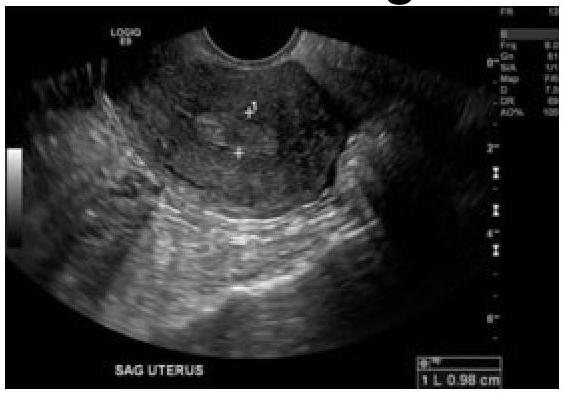
60-year-old female with a history of intermittent bleeding. What is the diagnosis based on the ultrasound image?
A sexually active female presenting with profuse frothy foul-smelling discharge with intense itching. Strawberry cervix revealed on examination. What will be the diagnosis?
45 years female with 3 months of menorrhagia. USG showing 2 cm submucosal fibroid. What is the most appropriate management option?
Practice by Chapter
Abnormal Uterine Bleeding
Practice Questions
Endometriosis
Practice Questions
Adenomyosis
Practice Questions
Uterine Fibroids
Practice Questions
Ovarian Cysts
Practice Questions
Pelvic Inflammatory Disease
Practice Questions
Vulvovaginitis
Practice Questions
Pelvic Organ Prolapse
Practice Questions
Vulvar Disorders
Practice Questions
Benign Breast Diseases
Practice Questions
Want unlimited practice?
Get full access to all questions, explanations, and performance tracking.
Scan to download app