
Gynecological Disorders — MCQs

On this page
MC site of injury to the ureter during a hysterectomy is:
Compared with other mullerian duct defects, a transverse vaginal septum is associated with lower rate of
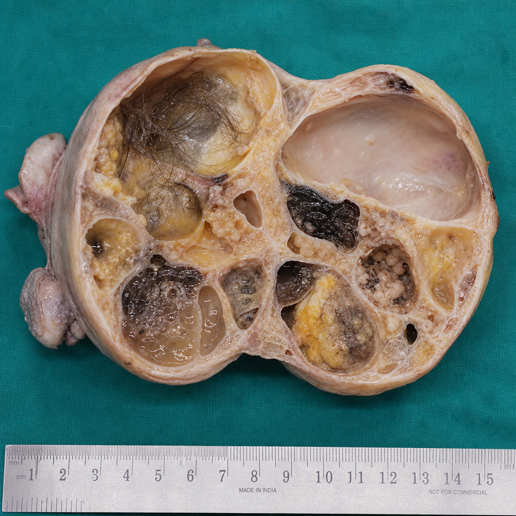
A 25-year-old female patient presented with complaints of feeling of mass per abdomen. On examination, a mass was found on left adnexal region with multiple solid cystic areas. The patient was operated and the mass was removed, which has been shown below. What is the true statement regarding the condition?
Distending media used in hysteroscopy are all except
To minimize ureteric damage, the following preoperative and operative precautions may be taken except:
Vaginal adenosis is evident in women who had exposure to which of the following in utero?
Most common complication of vaginoplasty is
All of the following are true about the isthmus EXCEPT:
Treatment of choice in a perimenopausal woman with bleeding PV due to multiple fibroids is:
A multiparous woman with a history of LSCS presents with cyclical hematuria and normal menstruation. The most likely diagnosis is:
Practice by Chapter
Abnormal Uterine Bleeding
Practice Questions
Endometriosis
Practice Questions
Adenomyosis
Practice Questions
Uterine Fibroids
Practice Questions
Ovarian Cysts
Practice Questions
Pelvic Inflammatory Disease
Practice Questions
Vulvovaginitis
Practice Questions
Pelvic Organ Prolapse
Practice Questions
Vulvar Disorders
Practice Questions
Benign Breast Diseases
Practice Questions
Want unlimited practice?
Get full access to all questions, explanations, and performance tracking.
Scan to download app