
Gynecological Disorders — MCQs

On this page
A patient complained of whitish discharge from the vagina and yellow staining on their clothes. There is no itching, no redness, and pH is acidic. What is the likely cause?
A woman presents with painless ulcers on the vulva, she gives a history of having multiple sexual partners and has had a stillbirth at 28 weeks in the past. What is the next best step of investigation?
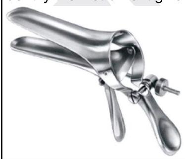
Identify the instrument given below.
A female patient having frothy vaginal discharge was found to have a strawberry cervix. Which of the following is the drug of choice?
Which of the following symptoms are seen in endometriosis? 1. Infertility 2. Dysmenorrhea 3. Vaginal discharge 4. Vaginal bleeding
A 24-year-old woman presents with abnormal vaginal discharge. Wet mount shows motile trichomonads. Her male partner is asymptomatic. Which of the following is the most appropriate management for her partner?
Which of the following is NOT a criterion in Amsel's criteria for diagnosing bacterial vaginosis?
A 25-year-old woman presents with thin, gray-white vaginal discharge and mild itching. Microscopy shows clue cells and pH is 5.5. What is the most appropriate treatment?
A 25 year old lady presented with curdy white discharge from the vagina is likely to be suffering from:-
Amsel's criteria are used for?
Practice by Chapter
Abnormal Uterine Bleeding
Practice Questions
Endometriosis
Practice Questions
Adenomyosis
Practice Questions
Uterine Fibroids
Practice Questions
Ovarian Cysts
Practice Questions
Pelvic Inflammatory Disease
Practice Questions
Vulvovaginitis
Practice Questions
Pelvic Organ Prolapse
Practice Questions
Vulvar Disorders
Practice Questions
Benign Breast Diseases
Practice Questions
Want unlimited practice?
Get full access to all questions, explanations, and performance tracking.
Scan to download app