
Gynecological Disorders — MCQs

On this page
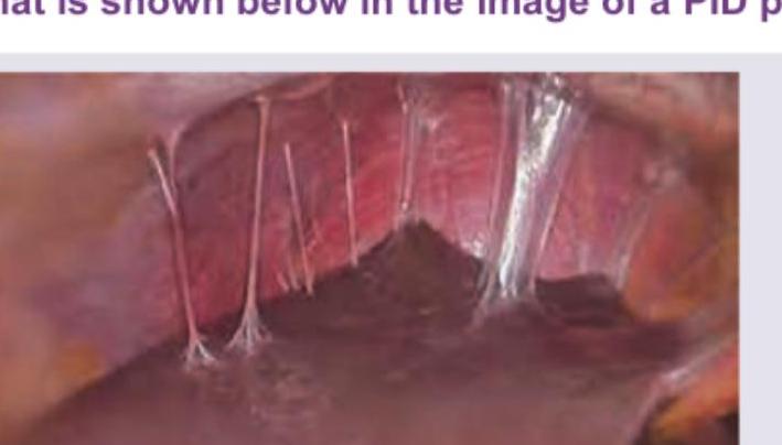
What is shown below in the image of a PID patient?
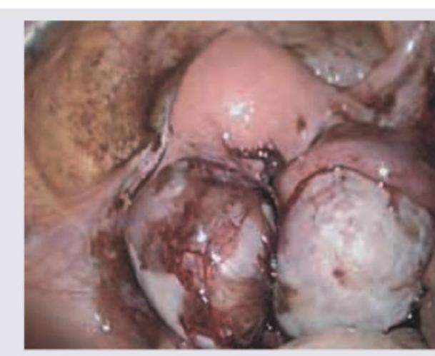
A 28 -year-old female patient presented with lower abdominal pain along with dysmenorrhea. The following finding was seen on laparoscopic examination. What is the likely diagnosis?
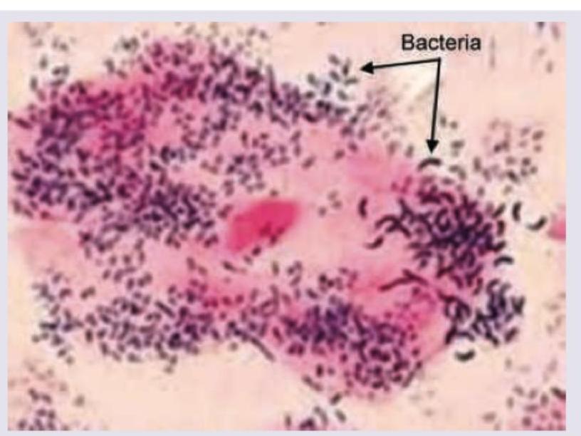
A 21-year-old girl is having vaginal discharge one week after unprotected sexual intercourse. The smear of vaginal discharge is shown below. All are diagnostic criteria for the condition shown except:
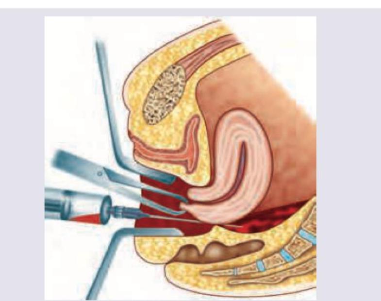
Which of the following is used in the gynecological procedure being performed in the image?
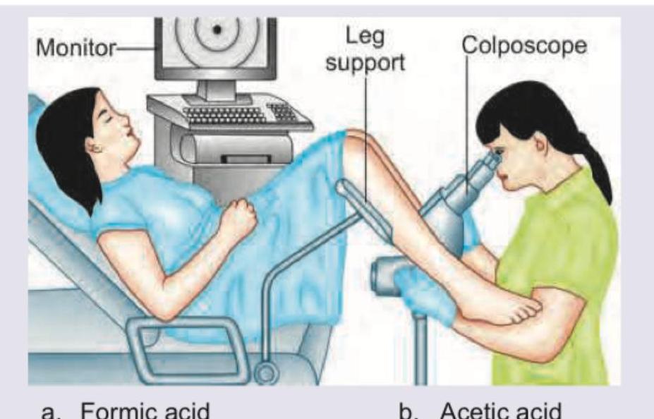
The procedure being performed is called:
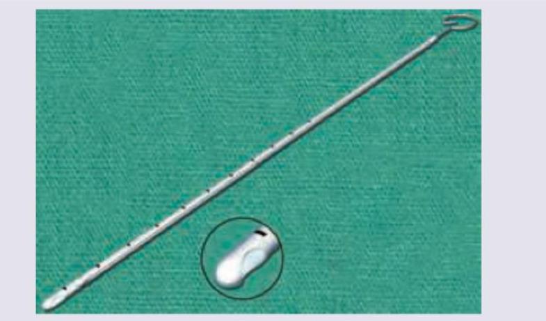
The image shows: (Recent Neet Pattern 2016-17)

The image shows: (Recent Neet Pattern 2016-17)
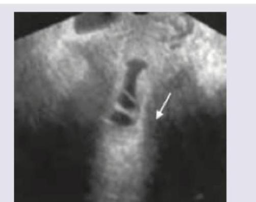
A patient presents with the following clinical finding on pelvic examination. What is the most likely diagnosis?
What is the best treatment for the condition shown below?
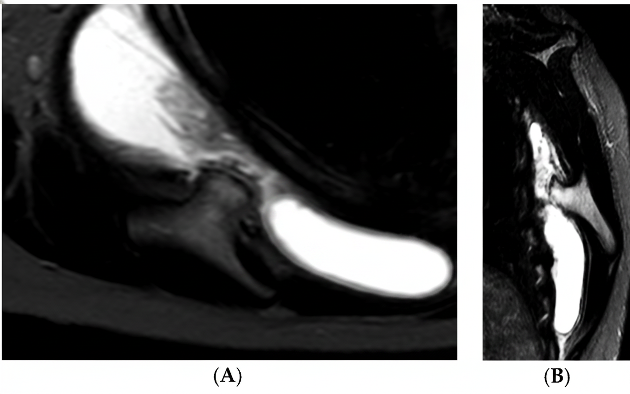
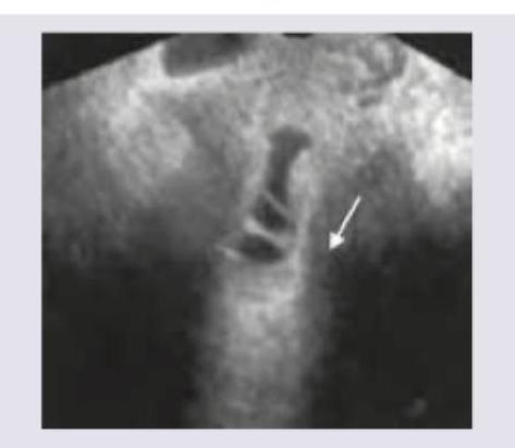
Transvaginal saline infusion sonographic evaluation shows:
Practice by Chapter
Abnormal Uterine Bleeding
Practice Questions
Endometriosis
Practice Questions
Adenomyosis
Practice Questions
Uterine Fibroids
Practice Questions
Ovarian Cysts
Practice Questions
Pelvic Inflammatory Disease
Practice Questions
Vulvovaginitis
Practice Questions
Pelvic Organ Prolapse
Practice Questions
Vulvar Disorders
Practice Questions
Benign Breast Diseases
Practice Questions
Want unlimited practice?
Get full access to all questions, explanations, and performance tracking.
Scan to download app