
Gynecological Disorders — MCQs

On this page
What is the treatment of choice for bacterial vaginosis?
Powder-burr appearance on laparoscopy is characteristic of:
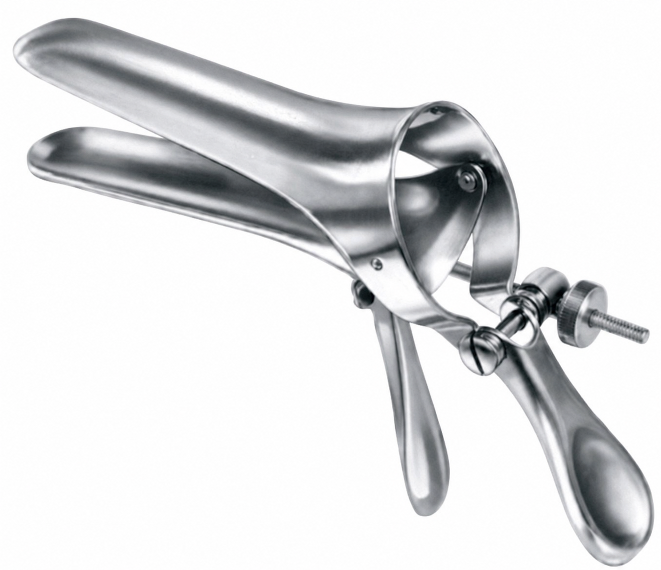
Identify the gynecological instrument shown in the image below:
A 36-year-old P2L2 patient diagnosed with severe endometriosis shows pelvic adhesions on laparoscopy. She has undergone tubal ligation and adhesiolysis previously. What is the most appropriate management during laparoscopy?
What is the best diagnostic test for evaluating endometrial pathology?
A 38 year old woman presents with complaints of heavy menstrual bleeding, pelvic discomfort, and frequent urination. On physical examination, her uterus is found to be irregularly enlarged. Which of the following is the most likely diagnosis?
A postmenopausal woman presents with irregular bleeding, endometrium biopsy shows endometrial hyperplasia without atypia. What is the likely management?
75% of iatrogenic ureteric injuries are due to gynaecological procedures. Which hysterectomy route has the least risk of ureteric injury?
The ureter is safe in which type of hysterectomy?
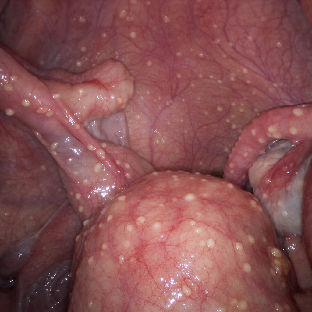
During laparoscopy, multiple small, yellowish-white tubercles are observed scattered over the pelvic peritoneum and adnexal surfaces. What is the most likely diagnosis?
Practice by Chapter
Abnormal Uterine Bleeding
Practice Questions
Endometriosis
Practice Questions
Adenomyosis
Practice Questions
Uterine Fibroids
Practice Questions
Ovarian Cysts
Practice Questions
Pelvic Inflammatory Disease
Practice Questions
Vulvovaginitis
Practice Questions
Pelvic Organ Prolapse
Practice Questions
Vulvar Disorders
Practice Questions
Benign Breast Diseases
Practice Questions
Want unlimited practice?
Get full access to all questions, explanations, and performance tracking.
Scan to download app