
Gynecological Disorders — MCQs

On this page
Pain in endometriosis is due to:
What is the most common symptom present in an undisturbed ectopic pregnancy?
Danazol used in the treatment of endometriosis causes which of the following changes within the endometrium and endometriosis tissue?
What is the cause of acidic pH of the vagina?
Endometrial hyperplasia is typically associated with which of the following conditions?
Which of the following is NOT true about fibroids?
Which of the following is NOT a concern when using mifepristone for the treatment of uterine fibroid?
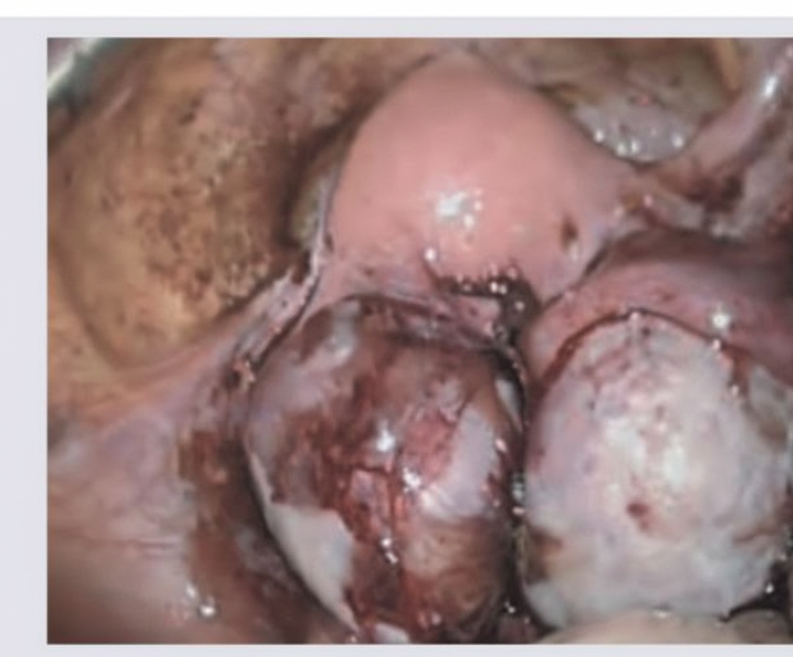
In the following laparoscopic view of the uterus and pelvis, what is the likely diagnosis?
A 34-year-old woman has been trying to conceive for 3 years. On pelvic examination, there is a nodular, tender uterosacral ligament, a retroverted but normal-sized uterus, and a right adnexal mass. A recent pelvic ultrasound reveals a 6-cm right complex ovarian mass. Her CA-125 is elevated. What is the initial next step in management?
Ampullary pregnancy ruptures generally at how many weeks?
Practice by Chapter
Abnormal Uterine Bleeding
Practice Questions
Endometriosis
Practice Questions
Adenomyosis
Practice Questions
Uterine Fibroids
Practice Questions
Ovarian Cysts
Practice Questions
Pelvic Inflammatory Disease
Practice Questions
Vulvovaginitis
Practice Questions
Pelvic Organ Prolapse
Practice Questions
Vulvar Disorders
Practice Questions
Benign Breast Diseases
Practice Questions
Want unlimited practice?
Get full access to all questions, explanations, and performance tracking.
Scan to download app