
Gynecological Disorders — MCQs

On this page
Which of the following is true regarding pain in endometriosis?
A young lady presents with a complaint of copious vaginal discharge. On per speculum examination, a 'strawberry vagina' is noted, with no cervical discharge. Which of the following should be given for management?
What is the definition of a unicornuate uterus?
Which of the following is NOT characteristic of bacterial vaginosis?
What is the best endometrial reaction in ectopic pregnancy?
Which of the following is true about a complete hydatidiform mole?
What is the best investigation for endometriosis?
What is the most common cause of vaginal discharge among reproductive-age females?
Hysteroscopic resection is indicated for submucosal myomas of which size?
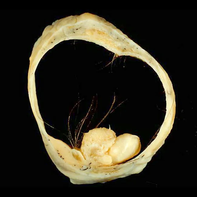
Which of the following statements is false regarding the given specimen?
Practice by Chapter
Abnormal Uterine Bleeding
Practice Questions
Endometriosis
Practice Questions
Adenomyosis
Practice Questions
Uterine Fibroids
Practice Questions
Ovarian Cysts
Practice Questions
Pelvic Inflammatory Disease
Practice Questions
Vulvovaginitis
Practice Questions
Pelvic Organ Prolapse
Practice Questions
Vulvar Disorders
Practice Questions
Benign Breast Diseases
Practice Questions
Want unlimited practice?
Get full access to all questions, explanations, and performance tracking.
Scan to download app