
Gynecological Disorders — MCQs

On this page
Which of the following is the causative organism for Bartholin's cyst?
What is the ideal treatment for a 55-year-old female with endometrial hyperplasia with atypia?
Which organ is NOT involved by endometriosis?
Which of the following is not a treatment option for a 29-year-old patient with moderate to severe endometriosis?
Regarding adenomyosis, all of the following statements are true EXCEPT:
Which of the following statements about uterine fibroids is FALSE?
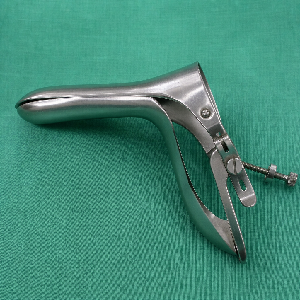
What is the name of the following instrument?
A 30-year-old female presents with chronic cyclical abdominal pain, which is increased during the 5 days of her menstrual cycle. The patient has been married for 2 years and has not conceived. What is the most appropriate next step in management?
A patient presents with greenish discharge and a "strawberry cervix." This clinical presentation is most commonly associated with which infection?
Hidradenoma of the vulva arises from which structure?
Practice by Chapter
Abnormal Uterine Bleeding
Practice Questions
Endometriosis
Practice Questions
Adenomyosis
Practice Questions
Uterine Fibroids
Practice Questions
Ovarian Cysts
Practice Questions
Pelvic Inflammatory Disease
Practice Questions
Vulvovaginitis
Practice Questions
Pelvic Organ Prolapse
Practice Questions
Vulvar Disorders
Practice Questions
Benign Breast Diseases
Practice Questions
Want unlimited practice?
Get full access to all questions, explanations, and performance tracking.
Scan to download app