Where are Gartner's cysts typically located?
What factor is associated with decreased success in the medical management of ectopic pregnancy?
What is the first-line investigation of choice for diagnosis of PID?
In your STI clinic, standardized treatment kits are available for different conditions based on clinical presentation and likely pathogens. A 22-year-old female comes to the STI clinic with minimal vaginal discharge. On speculum examination, erosions are seen on the cervix. Which of the following treatment kit should be given to this patient?
A woman comes with complaints of pain and swelling in the perineal area. She also has complaints of difficulty in walking and sitting. She gives a history of multiple sexual partners. On examination, a tender swelling is seen with redness on the labia. What is the most likely diagnosis?
A 16-year-old girl presents with cyclical pelvic pain every month. She has not achieved menarche yet. On examination, a suprapubic bulge can be seen in the lower abdomen. PR examination reveals a bulging swelling in the anterior aspect. What is the most likely diagnosis?
What is the best treatment option for a septate uterus?
Identify the type of hymen.

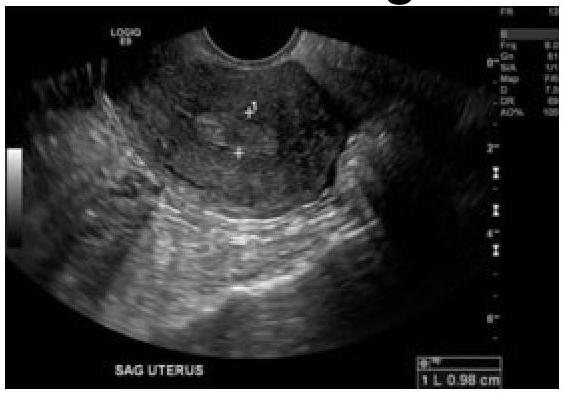
60-year-old female with a history of intermittent bleeding. What is the diagnosis based on the ultrasound image?

A sexually active female presenting with profuse frothy foul-smelling discharge with intense itching. Strawberry cervix revealed on examination. What will be the diagnosis?
Explanation: ***Anterolateral vaginal wall*** - **Gartner's cysts** are remnants of the **mesonephric (Wolffian) duct**. - These ducts primarily run along the **anterolateral vaginal wall**, which is their typical location. *Anterolateral cervix* - While located in the gynecological tract, the **cervix** is not the primary site for remnants of the **mesonephric duct**. - Cysts in this area are more commonly **Nabothian cysts**, which are retention cysts of cervical glands. *Posterolateral vaginal wall* - The **mesonephric duct** remnants, from which Gartner's cysts arise, typically course along the **anterior** rather than the posterior vaginal wall. - Cysts in the posterolateral wall are more likely to be **inclusion cysts** or related to the **Bartholin's glands**. *Posterolateral cervix* - Similar to the anterolateral cervix, this location is not typical for **Gartner's cysts**. - Cysts found here are likely to have a different embryological origin or pathological basis.
Explanation: ***Beta HCG > 5000 IU/L*** - A **beta-hCG level greater than 5000 IU/L** is a well-established **relative contraindication** for medical management with methotrexate. - High beta-hCG levels indicate a **larger ectopic mass with higher metabolic activity**, which significantly increases the risk of **treatment failure** and need for surgical intervention. - Most protocols recommend **surgical management** when beta-hCG exceeds 5000 IU/L due to decreased success rates with methotrexate. *Gestational sac > 3cm* - While a **large ectopic mass** (typically > 3.5-4 cm) is associated with lower success rates, the cutoff varies across guidelines. - Ectopic mass size is a relative contraindication, but **beta-hCG level** is a more standardized and objectively measurable criterion used in clinical protocols. *Duration of gestation > 5 weeks* - The **duration of gestation alone** is not a primary determinant of medical management success. - Other factors like **beta-hCG levels, mass size, and cardiac activity** are more critical in determining suitability for medical management. *Cardiac activity absent* - **Absence of cardiac activity** is actually a **favorable prognostic factor** for medical management, indicating a non-viable pregnancy with lower metabolic activity. - Medical management is often **contraindicated** when **fetal cardiac activity is present**, as it indicates a more viable and metabolically active pregnancy with higher failure risk.
Explanation: ***Ultrasonography*** - **Transvaginal ultrasonography (TVUS)** is the **first-line imaging investigation of choice** for diagnosing PID because it is **non-invasive**, readily available, cost-effective, and can visualize important diagnostic features such as adnexal masses, hydrosalpinx, free fluid, or tubo-ovarian abscesses. - While laparoscopy offers direct visualization, TVUS provides valuable diagnostic information with less risk and discomfort, making it the preferred initial investigation that guides further management. *Laparoscopy* - **Laparoscopy** is considered the **gold standard** for definitive diagnosis of PID as it allows direct visualization of the pelvic organs and can confirm inflammation, adhesions, or abscesses. - However, it is an **invasive surgical procedure** with associated risks and is therefore reserved for cases where the diagnosis is uncertain, when conservative management fails, or when complications are suspected. - It is not the first-line investigation due to its invasiveness and the need for anesthesia. *Colposcopy* - **Colposcopy** is a procedure used to **examine the cervix and vagina** with magnification; it is primarily used for screening and diagnosis of **cervical abnormalities** or cervical intraepithelial neoplasia. - It does not visualize the internal pelvic organs (uterus, fallopian tubes, ovaries) and therefore has no role in the diagnosis of PID. *Hysteroscopy* - **Hysteroscopy** involves inserting a scope into the **uterine cavity to visualize the endometrium** and inspect for intrauterine pathology such as polyps, fibroids, or adhesions. - While it can diagnose some uterine conditions, it does not allow for visualization of the fallopian tubes or adnexa, making it unsuitable for diagnosing PID which primarily affects these extrauterine structures.
Explanation: ***Grey*** - A grey kit is indicated for **vaginal discharge** with associated **cervical erosions**, suggesting a treatable bacterial STI like gonorrhea or chlamydia. This kit typically contains antibiotics effective against these pathogens. - The presence of **minimal vaginal discharge** combined with **cervical erosions** points towards cervicitis, for which the grey kit is specifically designed. *Green* - The green kit is typically for the treatment of **vaginal discharge** without specific signs of cervicitis, often targeting common causes like **bacterial vaginosis** or **trichomoniasis**. - It would not specifically address the **cervical erosions** seen in this patient, which are more indicative of cervicitis. *Red* - The red kit is generally used for the treatment of **genital ulcers**, which are typically caused by **herpes simplex virus** or **syphilis**. - While there are erosions, the primary complaint is discharge, and erosions are not typically the sole indicator for a "genital ulcer" kit. *Yellow* - The yellow kit is often designated for **urethral discharge** in males, addressing conditions like **gonorrhea** or **chlamydia** when presenting as urethritis. - This patient is female and presents with **vaginal discharge** and **cervical erosions**, making the yellow kit inappropriate.
Explanation: ***Bartholin abscess*** - The presentation of **painful, tender swelling with redness** on the labia, especially causing difficulty in walking and sitting, is classic for a **Bartholin gland abscess**. - **Multiple sexual partners** can increase the risk of infection leading to abscess formation due to bacterial contamination. *Chlamydial infection* - While Chlamydia is a sexually transmitted infection, it typically manifests as **cervicitis**, **urethritis**, or pelvic inflammatory disease, often with a discharge. - It does not directly cause an acute, localized labial abscess as described. *Genital Tuberculosis* - Genital tuberculosis is a **chronic condition** that usually presents with menstrual irregularities, infertility, or chronic pelvic pain. - It rarely causes an acute, tender labial swelling or abscess. *Herpes infection* - Genital herpes presents with **painful vesicles or ulcers** on the genitalia, often associated with a prodrome of itching or tingling. - It does not typically cause a single, large, tender, and red fluctuant swelling indicative of an abscess.
Explanation: ***Imperforate hymen*** - The combination of **cyclical pelvic pain** without menarche (primary amenorrhea) and a **suprapubic bulge** with **bulging swelling on PR examination** strongly suggests an imperforate hymen. - This condition leads to the **accumulation of menstrual blood (hematocolpos)**, causing the observed swelling and pain. - Imperforate hymen is the **most distal obstruction** of the female genital tract, presenting with a characteristic **bulging membrane at the vaginal opening**. *Transverse vaginal septum* - This condition also causes **primary amenorrhea** and **hematocolpos** leading to cyclical pain. - However, a transverse vaginal septum is located **higher in the vagina** (not at the introitus) and would not typically present with such an obvious **bulging swelling on examination** at the vaginal opening. *Vaginal atresia* - **Vaginal atresia** involves the complete or partial absence of the vagina, which would prevent menarche and cause cyclical pain. - While it results in hematocolpos (if the uterus is present), the presentation differs from the classic **bulging membrane** seen with imperforate hymen. *Cervical agenesis* - **Cervical agenesis** is the congenital absence or incomplete formation of the cervix, leading to **primary amenorrhea** and severe cyclical pain due to retained menstrual blood in the uterus (**hematometra**). - This condition would not present with a **bulging mass on PR examination** at the vaginal level, but rather with an enlarged uterus above, as the obstruction is at the cervical level, not at the vaginal outlet.
Explanation: ***Transcervical hysteroscopic resection of the septum*** - This procedure involves using a **hysteroscope** to visualize and resect the **fibrous or muscular septum** that divides the uterine cavity, restoring a normal uterine shape. - It is considered the gold standard due to its **minimally invasive nature**, effectiveness in improving reproductive outcomes, and lower risk of complications compared to abdominal approaches. *Tompkins Metroplasty* - This is an **abdominal surgical procedure** primarily used for the repair of a **bicornuate uterus**, not typically for a septate uterus. - It involves resecting the uterine fundus to create a single uterine cavity, which is more invasive than hysteroscopic septum resection. *Jones metroplasty* - This procedure is also an **abdominal approach** used for the surgical correction of a **bicornuate uterus**, not a septate uterus. - It involves excising the septal portion and approximating the uterine walls. *Strassmann metroplasty* - This is another **abdominal surgical technique** that is primarily indicated for the repair of a **bicornuate or didelphys uterus**, where a large defect needs to be corrected. - It involves reconstructing the uterus through a fundal incision, which is significantly more invasive than hysteroscopic septal resection for a septate uterus.
Explanation: ***Septate hymen*** - The image clearly displays a **band of tissue** running across the hymenal opening, dividing it into two smaller openings, which is characteristic of a **septate hymen**. - This type of hymen can sometimes interfere with menstruation or intercourse and may require surgical correction. *Imperforate hymen* - An **imperforate hymen** completely covers the vaginal opening, with no perforations for menstrual flow. - This typically presents at menarche with **abdominal pain** and a bulging hymen due to retained menstrual blood (hematocolpos). *Semilunar hymen* - A **semilunar hymen** is incomplete, forming a crescent shape at the posterior aspect of the vaginal opening. - This is a common and normal variant, usually not causing any clinical issues. *Annular hymen* - An **annular hymen** is characterized by a circular opening in the center, encircled by hymenal tissue, appearing like a ring. - This is a common hymenal configuration and usually allows for normal menstrual flow.
Explanation: ***Endometrial polyp*** - The ultrasound image shows a well-defined, **echogenic focal lesion within the endometrial cavity**, which is characteristic of an endometrial polyp. - In a 60-year-old female with intermittent bleeding, a polyp is a common cause of **postmenopausal bleeding** and is often identified as an intracavitary mass on ultrasound. - Endometrial polyps appear as **focal, hyperechoic or isoechoic masses** with a smooth contour projecting into the endometrial cavity. *Ca endometrium* - **Endometrial carcinoma** typically appears as diffuse endometrial thickening (>4-5 mm in postmenopausal women), irregular endometrial-myometrial interface, or heterogeneous endometrial echoes, often with evidence of myometrial invasion. - While bleeding is a cardinal symptom of endometrial carcinoma, the distinct **focal, well-circumscribed nature** of the lesion on ultrasound is less typical for carcinoma, which tends to be more diffuse and irregular. *Submucosal fibroid* - A **submucosal fibroid** (leiomyoma) is a benign smooth muscle tumor originating from the myometrium that protrudes into the endometrial cavity. - While it can appear as an intracavitary mass and cause abnormal bleeding, fibroids often have a more **heterogeneous or hypoechoic appearance** with posterior acoustic shadowing, compared to the isoechoic to hyperechoic appearance of a polyp. - The feeding vessel sign (blood flow at the base) is more characteristic of polyps than fibroids. *Endometrial hyperplasia* - **Endometrial hyperplasia** presents as **diffuse, uniform endometrial thickening** rather than a focal intracavitary mass. - It typically shows homogeneous increased echogenicity of the entire endometrium without a discrete, well-defined lesion as seen with a polyp. - While it can cause postmenopausal bleeding, the focal nature of the lesion in this case makes hyperplasia less likely.
Explanation: **Trichomonas vaginalis** - The classic presentation of **profuse, frothy, foul-smelling vaginal discharge** with **intense itching** and the presence of a **strawberry cervix** are highly characteristic of a *Trichomonas vaginalis* infection. - *Trichomonas vaginalis* is a **motile protozoan** and a common sexually transmitted infection. *Bacterial vaginosis* - While bacterial vaginosis (BV) causes a **foul-smelling discharge** (often described as "fishy"), it is typically **thin and grayish-white**, not frothy, and does not cause a strawberry cervix. - BV is caused by an **overgrowth of anaerobic bacteria** and a decrease in lactobacilli, leading to an elevated vaginal pH. *Candidiasis* - Candidiasis (yeast infection) presents with a **thick, white, "cottage cheese-like" discharge** and intense itching, but the discharge is usually odorless and it does not cause a frothy discharge or strawberry cervix. - It is caused by an **overgrowth of *Candida* species**, primarily *Candida albicans*, and is not typically sexually transmitted. *None of the options* - Given the classic constellation of symptoms and definitive signs, a specific diagnosis can be made, making this option incorrect.
Abnormal Uterine Bleeding
Practice Questions
Endometriosis
Practice Questions
Adenomyosis
Practice Questions
Uterine Fibroids
Practice Questions
Ovarian Cysts
Practice Questions
Pelvic Inflammatory Disease
Practice Questions
Vulvovaginitis
Practice Questions
Pelvic Organ Prolapse
Practice Questions
Vulvar Disorders
Practice Questions
Benign Breast Diseases
Practice Questions
Get full access to all questions, explanations, and performance tracking.
Start For Free