A 55-year-old woman presents with postmenopausal bleeding. An endometrial biopsy reveals atypical endometrial hyperplasia. What is the most appropriate treatment?
A 30-year-old woman presents with secondary amenorrhea and a history of curettage. What is the diagnosis?
A 34-year-old woman presents with pelvic pain and a palpable adnexal mass. The CA-125 level is elevated. What is the next best step in management?
A 30-year-old woman presents with vaginal discharge and pelvic pain. Examination reveals cervical motion tenderness. What is the next step?
What is the most reliable method to confirm the placement of a ring pessary in a patient with uterine prolapse?
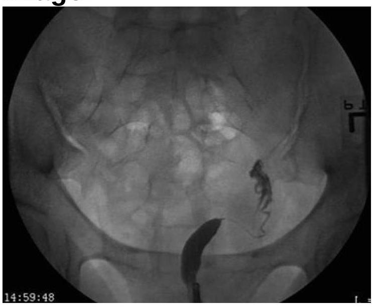
Based on the provided image, which of the following is the correct diagnosis?

Which of the following is NOT included in the Nugent score?
Kamla, a 30-year-old woman, P2L2 with a 3.2 x 4.1 cm fibroid uterus, presents with menorrhagia and has been on symptomatic treatment for the past 6 months. She refuses surgery. What is the next line of management?
What is the definitive treatment for adenomyosis?
Which of the following best describes endometriosis?
Explanation: ***Hysterectomy*** - **Atypical endometrial hyperplasia** carries a significant risk (25-50%) of progression to **endometrial cancer**, especially in postmenopausal women. - **Hysterectomy** (removal of the uterus) is the most appropriate definitive treatment to eliminate the hyperplastic tissue and prevent future malignant transformation. *Observation* - **Observation** is not recommended for atypical hyperplasia due to the high risk of **malignant progression**. - It would be considered only for simple or complex hyperplasia without atypia, or in very specific cases where surgery is contraindicated. *Progesterone therapy* - **Progesterone therapy** can be used to treat simple or complex hyperplasia without atypia, or atypical hyperplasia in women who desire future fertility. - However, for postmenopausal women with **atypical hyperplasia**, the efficacy of medical management is lower, and the risk of progression often warrants surgical intervention. *Endometrial ablation* - **Endometrial ablation** destroys the endometrial lining and is primarily used to treat dysfunctional uterine bleeding. - It is **contraindicated** in cases of atypical hyperplasia or malignancy because it can obscure the diagnosis and prevent adequate sampling if cancer is present.
Explanation: ***Asherman syndrome*** - **Asherman syndrome** is characterized by the formation of **intrauterine adhesions** (scar tissue), typically as a complication of uterine surgeries like **curettage**. - These adhesions can lead to **secondary amenorrhea** by preventing normal endometrial growth and shedding, despite normal ovarian function. *Polycystic ovary syndrome* - **PCOS** presents with **oligomenorrhea** or amenorrhea, but it's usually primary or secondary amenorrhea without a clear history of uterine instrumentation. - Key features include **hyperandrogenism** (hirsutism, acne) and **polycystic ovaries** on ultrasound, which are not mentioned. *Hypothyroidism* - **Hypothyroidism** can cause menstrual irregularities, including **amenorrhea**, due to its impact on the hypothalamic-pituitary-gonadal axis. - However, it typically presents with other systemic symptoms like **fatigue**, **weight gain**, and **cold intolerance**, and the history of curettage directly points to a uterine cause. *Premature ovarian failure* - **Premature ovarian failure (POF)** involves the cessation of ovarian function before age 40, leading to **amenorrhea** and menopausal symptoms like hot flashes and vaginal dryness. - It would be associated with **elevated FSH** and **low estrogen**, but the history of curettage makes Asherman syndrome a more direct explanation for secondary amenorrhea.
Explanation: ***Transvaginal ultrasound*** - A **transvaginal ultrasound** is the initial and most appropriate imaging modality for evaluating a **palpable adnexal mass** and determining its characteristics. - It helps distinguish between benign and malignant features, guiding further diagnostic and management steps before more invasive procedures. *Immediate laparotomy* - **Immediate laparotomy** is an invasive surgical procedure and is typically reserved for cases with a high suspicion of malignancy or acute emergencies, such as **ruptured ectopic pregnancy** or **ovarian torsion**, after initial imaging and diagnostic workup. - Performing a laparotomy without prior imaging for characterization of the mass could lead to unnecessary surgery if the mass is benign or could be managed less invasively. *CT scan of the abdomen and pelvis* - While a **CT scan** provides detailed anatomical information, it is generally considered a secondary imaging study for ovarian masses and is performed after an initial ultrasound has characterized the mass. - CT scans expose the patient to **ionizing radiation** and are less effective than ultrasound for detailed characterization of ovarian mass morphology. *MRI of the pelvis* - An **MRI of the pelvis** offers excellent soft-tissue contrast and can further characterize indeterminate adnexal masses found on ultrasound, often used to differentiate between benign and malignant lesions. - However, it is a more expensive and time-consuming imaging modality and is typically performed after an initial **transvaginal ultrasound** if the findings are equivocal or require further clarification.
Explanation: ***Antibiotics*** - The symptoms of **vaginal discharge**, **pelvic pain**, and **cervical motion tenderness** are highly suggestive of **Pelvic Inflammatory Disease (PID)**. - Initial management for PID is typically prompt empirical antibiotic therapy to prevent complications such as infertility or ectopic pregnancy *Laparoscopy* - This is a more invasive procedure generally used for definitive diagnosis or management of complications like **tubo-ovarian abscesses** or when the diagnosis is uncertain despite initial treatment. - It is not the **first-line intervention** for suspected PID without failed medical management or red flag symptoms. *Ultrasound* - While ultrasound can be helpful in identifying complications of PID, such as **tubo-ovarian abscesses** or ruling out other conditions. - It is not the **initial therapeutic step** for suspected PID, where timely antibiotic treatment is crucial. *Hysteroscopy* - This procedure involves inserting a thin scope into the uterus to visualize the uterine cavity. - It is primarily used for investigating causes of abnormal uterine bleeding, infertility, or recurrent miscarriage, and is **not indicated** for the initial management of PID.
Explanation: ***Pelvic examination*** - A **pelvic examination** allows direct visualization and palpation of the pessary to ensure it is correctly seated in the posterior vaginal fornix and behind the pubic symphysis, providing proper support. - This method enables the clinician to assess for proper fit, ensuring it is not too tight or too loose, and that it does not cause **discomfort** or **vaginal wall erosion**. - This is the **gold standard** for confirming correct pessary placement. *Patient reports relief of prolapse symptoms* - While symptom relief is a desirable outcome, it is a **subjective measure** and does not directly confirm the anatomical correctness of pessary placement. - Initial symptom relief might occur even with suboptimal placement, which could lead to later complications such as **pessary displacement** or **vaginal irritation**. *Absence of vaginal discharge* - The absence of vaginal discharge is not a reliable indicator because **vaginal discharge** can be present or absent for various reasons unrelated to pessary placement. - Some women may experience an increase in physiological discharge or develop infections even with a properly placed pessary, while others may not experience discharge even with improper placement. *Absence of pain* - Lack of immediate pain does not guarantee proper placement; a pessary can be improperly placed without causing acute pain, especially if it is too loose. - Pain or discomfort may develop later, either during activity or due to **pressure on the vaginal walls** if the fit is incorrect, so immediate absence of pain is not definitive.
Explanation: ***Unicornuate Uterus*** - The image distinctly shows **only one fallopian tube and one rudimentary uterine horn** on the right side, indicating a unicornuate uterus. - This malformation results from the **incomplete development of one Müllerian duct**, leading to a single, banana-shaped uterine cavity. *Uterus didelphys* - This condition involves **two completely separate uteri**, each with its own cervix and vagina. - The image does not show evidence of two distinct uterine bodies or cervices. *Bicornuate Uterus* - A bicornuate uterus is characterized by **two uterine horns that fuse caudally**, creating a heart-shaped appearance with a shared cervix. - The image clearly lacks the characteristic heart shape and shows only one functional horn. *Septate uterus* - A septate uterus has a **fibrous or muscular septum** dividing the uterine cavity, while the external uterine contour remains normal. - The image does not show a septum or a normal external uterine contour with an internal division; instead, it presents with a single underdeveloped horn.
Explanation: ***Gonococcus*** - The **Nugent score** is a **microscopic grading system** used to diagnose **bacterial vaginosis**, not sexually transmitted infections caused by specific bacteria like *Gonococcus*. - *Gonococcus* (specifically *Neisseria gonorrhoeae*) is indicative of **gonorrhea**, a separate clinical entity requiring different diagnostic methods like NAAT or culture. *Lactobacillus* - The Nugent score assesses the **morphotypes of bacteria** present on a **Gram-stained vaginal smear**, and a high number of *Lactobacillus* species is associated with a **healthy vaginal flora**. - A score of **0-3** indicates normal flora, characterized by abundant lactobacilli and absence of other morphotypes. *Gardnerella* - The Nugent score assigns points based on the presence of **small, curved Gram-variable rods** (typically *Gardnerella* species or similar). - An increase in these **coccobacillary morphotypes** contributes to a higher score, indicating a shift towards bacterial vaginosis. *Mobiluncus* - **Curved Gram-variable rods**, specifically *Mobiluncus* species, are also assessed in the Nugent score. - Their presence contributes points towards a higher score, as they are commonly associated with the pathogenesis of **bacterial vaginosis**.
Explanation: ***GnRH analogs to temporarily reduce fibroid size and control symptoms.*** - GnRH analogs induce a **hypoestrogenic state**, leading to a significant (up to 50%) reduction in fibroid size and resolution of menorrhagia. - This is a suitable non-surgical option for patients who decline surgery and have failed symptomatic treatment, offering temporary relief and potentially improving their anemia before other definitive treatments. *Danazol for symptomatic relief of menorrhagia.* - Danazol is an **androgen derivative** that can reduce menorrhagia, but it has significant **androgenic side effects** (e.g., hirsutism, acne, weight gain) that make it less desirable for long-term use. - It does not directly shrink fibroids effectively and is generally reserved for patients who cannot tolerate other hormonal therapies. *Myomectomy for fibroid removal (surgical option).* - Myomectomy is a surgical procedure to remove fibroids while preserving the uterus, but the patient explicitly **refuses surgery**, making this an inappropriate immediate next step. - While it is a definitive treatment for fibroids causing menorrhagia, patient preference must be respected. *Uterine artery embolization (UAE) to shrink fibroids and control symptoms.* - UAE is an effective **minimally invasive procedure** to shrink fibroids by blocking their blood supply, but it is considered an interventional radiological procedure. - Although less invasive than myomectomy, it still involves an invasive procedure and the question implies exploring non-surgical *medical* management first, given the refusal of surgery.
Explanation: ***Hysterectomy*** - **Hysterectomy** (surgical removal of the uterus) is the only definitive treatment for adenomyosis as it removes the ectopic endometrial tissue embedded within the myometrium. - This procedure alleviates symptoms such as **heavy menstrual bleeding (menorrhagia)** and **severe pelvic pain** by eliminating the source of the problem. *OC pills* - **Oral contraceptive pills** can help manage the symptoms of adenomyosis, such as heavy bleeding and pain, by suppressing endometrial growth. - However, they do not remove the **ectopic endometrial tissue** and therefore are not a definitive cure for the condition. *NSAIDS* - **NSAIDs (Nonsteroidal Anti-Inflammatory Drugs)** are used for symptomatic relief of pain associated with adenomyosis, particularly dysmenorrhea. - They reduce **prostaglandin production** and inflammation, but they do not address the underlying pathology or provide a definitive cure. *Endometrial ablation* - **Endometrial ablation** destroys the uterine lining and is primarily used for abnormal uterine bleeding. - It is generally *not effective* for adenomyosis because the ectopic endometrial tissue is deep within the **myometrium**, beyond the reach of ablation.
Explanation: ***Endometrial-like tissue located outside the uterus.*** - Endometriosis is defined by the presence of **endometrial-like tissue** (glands and stroma) outside the uterine cavity. - This ectopic tissue responds to **hormonal fluctuations** of the menstrual cycle, leading to bleeding, inflammation, and pain. *Endometrial tissue found within the myometrium.* - This condition describes **adenomyosis**, where endometrial glands and stroma are invaginated into the **myometrium** (uterine muscle wall). - While also causing pain and heavy bleeding, it is distinct from endometriosis where the tissue is located *outside* the uterus. *Myometrial tissue found within the endometrium.* - Myometrial tissue is normally the muscle layer of the uterus; finding it within the endometrium (the inner lining) is not a described medical condition. - This statement generally signifies a **misunderstanding of uterine anatomy** and common pathologies. *A rare type of endometrial cancer.* - Endometriosis is a **benign condition**, although rarely it can undergo malignant transformation into **endometrioid adenocarcinoma** or clear cell carcinoma. - It is not itself a type of cancer, but rather an inflammatory and proliferative disease.
Abnormal Uterine Bleeding
Practice Questions
Endometriosis
Practice Questions
Adenomyosis
Practice Questions
Uterine Fibroids
Practice Questions
Ovarian Cysts
Practice Questions
Pelvic Inflammatory Disease
Practice Questions
Vulvovaginitis
Practice Questions
Pelvic Organ Prolapse
Practice Questions
Vulvar Disorders
Practice Questions
Benign Breast Diseases
Practice Questions
Get full access to all questions, explanations, and performance tracking.
Start For Free