
Gynecologic Oncology — MCQs

On this page
Acetic acid staining of the cervix shows the following findings EXCEPT:
Recurrence of hydatiform mole is assessed by:
A 40-year-old woman has CIN 2. What is the next step in management?
Which of the following would be the earliest symptom of Ca cervix?
Endometrial cancer is associated with all EXCEPT:
Recurrence of gestational trophoblastic tumor can be associated with all except?
Which is the commonest malignancy of the ovary?
Which of the following is a serum marker for ovarian carcinoma?
What is the management for stage IIB cancer of the cervix?
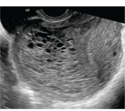
What is the diagnosis based on the ultrasound findings of an early pregnancy?
Practice by Chapter
Cervical Cancer
Practice Questions
Endometrial Cancer
Practice Questions
Ovarian Cancer
Practice Questions
Vulvar and Vaginal Cancer
Practice Questions
Gestational Trophoblastic Disease
Practice Questions
Screening for Gynecologic Cancers
Practice Questions
Principles of Gynecologic Oncology Surgery
Practice Questions
Radiation Therapy in Gynecologic Malignancies
Practice Questions
Chemotherapy in Gynecologic Oncology
Practice Questions
Palliative Care in Gynecologic Oncology
Practice Questions
Want unlimited practice?
Get full access to all questions, explanations, and performance tracking.
Scan to download app