Pain of ovarian carcinoma is typically referred to which anatomical region?
A 45-year-old woman presents to the gynecology department with prolonged vaginal bleeding. On examination, a cervical lesion is found that bleeds on touch. Her past history is insignificant. She had a Pap smear approximately 10 years ago, which was normal. What is the next best investigation?
What condition is characterized by a 'snow storm' appearance on ultrasound (IJSG)?
What is the estimated time taken for the conversion of CIN cervix to invasive carcinoma in years?
Which of the following is true about dysgerminoma of the ovary?
What is the chemotherapy regimen for dysgerminoma?
What is the most common ovarian malignancy in the post-menopausal period?
A 62-year-old postmenopausal woman presents with a blood-tinged vaginal discharge. Her last menstrual period was 10 years ago. On bimanual pelvic examination, the uterus is normal in size, with no palpable adnexal masses. There are no cervical erosions or masses. Her body mass index is 33. She has a 30-year history of hypertension and type 2 diabetes mellitus. An endometrial biopsy specimen is most likely to show which of the following?
Which of the following is NOT a common primary tumor of the vulva?
What is the first change observed on colposcopy in a precancerous lesion?
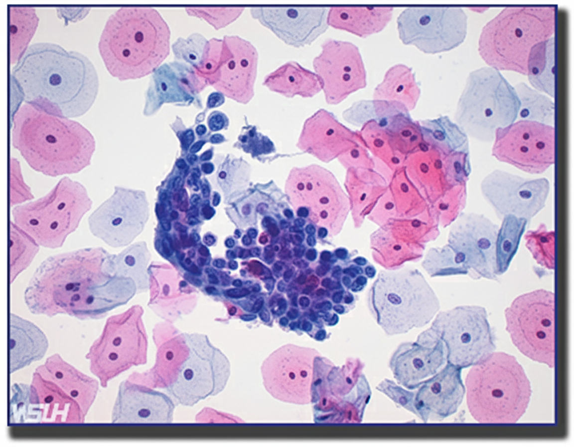
Explanation: **Explanation:** The referred pain of ovarian carcinoma to the **medial aspect of the thigh** is a classic anatomical correlation based on shared nerve supply. **1. Why the Medial Aspect of the Thigh is Correct:** The ovaries are embryologically derived from the same level as the kidneys (T10-T11), but their primary sensory innervation is closely associated with the **obturator nerve (L2-L4)**. When an ovarian mass or carcinoma enlarges, it can cause irritation or compression of the obturator nerve as it courses through the lateral pelvic wall. Since the obturator nerve provides sensory innervation to the skin of the **medial thigh**, pain is referred to this specific dermatomal distribution. **2. Analysis of Incorrect Options:** * **A. Posterior aspect of the thigh:** This area is supplied by the posterior cutaneous nerve of the thigh and the sciatic nerve. Pain here is more typical of lumbosacral disc herniation or pelvic masses compressing the sacral plexus. * **B. Cervical region:** This is anatomically unrelated. Referred pain to the shoulder/cervical region in gynecology usually suggests diaphragmatic irritation (e.g., hemoperitoneum from a ruptured ectopic pregnancy). * **C. Anterior aspect of the thigh:** This area is primarily supplied by the femoral nerve (L2-L4). While the nerve roots are similar, the specific clinical manifestation of ovarian pathology is classically linked to the obturator distribution. **3. Clinical Pearls for NEET-PG:** * **Obturator Sign:** Pain on internal rotation of the hip; though usually associated with appendicitis, it can be positive in pelvic inflammatory disease or ovarian masses. * **Howship-Romberg Sign:** Pain in the medial thigh exacerbated by hip extension/abduction, seen in obturator hernias (common in elderly, thin women). * **Lymphatic Spread:** Remember that ovarian cancer primarily spreads via **exfoliation** (peritoneal seeding), but its primary lymphatic drainage is to the **para-aortic lymph nodes** (at the level of the renal vessels).
Explanation: **Explanation:** The clinical presentation of a 45-year-old woman with **prolonged vaginal bleeding** and a **visible cervical lesion that bleeds on touch** is highly suspicious for **Cervical Carcinoma**. In any patient with a clinically visible growth or suspicious lesion on the cervix, the definitive next step is a **tissue diagnosis**. 1. **Why Cervical Punch Biopsy is correct:** A punch biopsy is the gold standard for diagnosing a visible cervical lesion. It allows for histological confirmation of malignancy and determines the cell type (Squamous cell vs. Adenocarcinoma). Note that if a lesion is visible, a Pap smear is unnecessary and may provide a false negative; direct biopsy is mandatory. 2. **Why other options are incorrect:** * **Hysteroscopy:** This is used to evaluate the uterine cavity (endometrium). While it helps in cases of abnormal uterine bleeding (AUB), it is not the primary investigation for a visible cervical growth. * **MRI Pelvis:** MRI is the investigation of choice for **staging** (evaluating tumor size and parametrial involvement) *after* malignancy has been histologically confirmed. It is not a diagnostic tool for the primary lesion. * **LEEP:** This is a diagnostic-cum-therapeutic procedure used for CIN (Cervical Intraepithelial Neoplasia) when the transformation zone is not fully visible or there is a discrepancy in cytology. It is not the initial step for a large, suspicious clinical growth. **Clinical Pearls for NEET-PG:** * **Most common symptom of Cervical Cancer:** Post-coital bleeding (though intermenstrual or prolonged bleeding is common). * **Screening vs. Diagnosis:** Pap smear/HPV DNA is for **screening** asymptomatic women; Biopsy is for **diagnosing** symptomatic women with a lesion. * **Staging:** Cervical cancer is staged **clinically** (FIGO staging), though the 2018 revision allows the use of imaging (MRI/CT) and pathology where available.
Explanation: **Explanation:** The **'snow storm' appearance** is the classic pathognomonic ultrasonographic finding of a **Hydatidiform Mole** (specifically a Complete Mole). This appearance is caused by the presence of multiple hydropic (swollen) chorionic villi and intrauterine hemorrhage. On ultrasound, these vesicles appear as multiple small echogenic foci interspersed with tiny cystic (anechoic) spaces, filling the entire endometrial cavity without a visible fetus or gestational sac. **Analysis of Options:** * **Hydatidiform Mole (Correct):** The proliferation of trophoblastic tissue leads to the characteristic vesicular pattern. In a Complete Mole (46,XX), there is no fetal tissue, whereas a Partial Mole may show a fetus with focal cystic changes in the placenta. * **Invasive Mole:** While it arises from a hydatidiform mole, it is characterized by the invasion of these vesicles into the **myometrium**. The ultrasound would show focal myometrial echoes and increased vascularity (on Doppler) rather than just a confined intrauterine 'snow storm.' * **Twin Pregnancy:** This would show two distinct gestational sacs or two fetuses with separate heartbeats. * **Ectopic Pregnancy:** Typically presents with an empty uterus, a pseudo-gestational sac, and an adnexal mass (e.g., 'blob sign' or 'tubal ring sign'). **High-Yield Clinical Pearls for NEET-PG:** * **hCG Levels:** Extremely high (often >100,000 mIU/mL) in complete moles. * **Ovarian Finding:** Bilateral **Theca Lutein Cysts** are often seen due to high hCG levels (hyperreactio luteinalis). * **Histopathology:** "Swiss cheese" appearance or "bunch of grapes" appearance macroscopically. * **Management:** Suction and evacuation is the treatment of choice, followed by weekly hCG monitoring until three consecutive negative results are obtained.
Explanation: **Explanation:** The progression of Cervical Intraepithelial Neoplasia (CIN) to invasive cervical carcinoma is a slow, multi-step process driven by persistent infection with high-risk Human Papillomavirus (HPV). **1. Why Option B (10 years) is correct:** On average, it takes approximately **10 to 15 years** for a high-grade CIN lesion (CIN 2 or 3) to progress to invasive squamous cell carcinoma. This long "latent period" or "pre-invasive phase" is the physiological basis for cervical cancer screening programs (like Pap smears and HPV DNA testing). Because the transition takes roughly a decade, regular screening can detect and treat cellular changes in the pre-malignant stage, effectively preventing the development of invasive cancer. **2. Why other options are incorrect:** * **Option A (5 years):** While rapid progression can occur in immunocompromised patients (e.g., those with HIV), it is not the standard timeline for the general population. * **Options C & D (15-20 years):** While some cases may take up to 20 years, "10 years" is the most frequently cited high-yield figure in standard textbooks (like Williams Gynecology and Shaw’s) for the average duration of progression from CIN to malignancy. **High-Yield Clinical Pearls for NEET-PG:** * **Most common site:** The Transformation Zone (Squamocolumnar junction). * **HPV Strains:** HPV 16 and 18 are responsible for ~70% of invasive cancers. * **Regression:** Not all CIN lesions progress; CIN 1 has a high rate of spontaneous regression (up to 60-80%), whereas CIN 3 has the highest risk of progression. * **Screening Goal:** The primary goal of screening is to identify **CIN 2 and CIN 3** (collectively termed high-grade squamous intraepithelial lesions or HSIL), as these are the immediate precursors to invasive cancer.
Explanation: **Explanation:** Dysgerminoma is the most common malignant germ cell tumor (GCT) of the ovary, typically affecting young women and adolescents. It is the female counterpart of the testicular seminoma. * **Option A (Blood spread):** Unlike most epithelial ovarian cancers which spread primarily via the transcoelomic route (exfoliation into the peritoneal cavity), dysgerminomas have a high propensity for **lymphatic spread** (to retroperitoneal nodes) and **hematogenous (blood) spread** to distant organs like the lungs and liver. * **Option B (Schiller-Duval bodies):** While Schiller-Duval bodies are the classic hallmark of **Yolk Sac Tumors** (Endodermal Sinus Tumors), they can occasionally be seen in mixed germ cell tumors involving a dysgerminoma component. However, in the context of this specific question and standard NEET-PG patterns, the examiner considers the presence of these bodies and the general characteristics of GCTs collectively. * **Option C (Radiosensitivity):** Dysgerminoma is unique among ovarian malignancies for being **exquisitely radiosensitive**. Although chemotherapy (BEP regimen) is now the preferred treatment to preserve fertility, radiotherapy remains a highly effective historical treatment modality. Since all three statements represent clinical or pathological characteristics associated with dysgerminoma or its management, **Option D** is the correct choice. **High-Yield Clinical Pearls for NEET-PG:** * **Tumor Marker:** LDH (Lactate Dehydrogenase) is the specific marker. It may also show mild elevations in hCG if syncytiotrophoblast giant cells are present. * **Microscopy:** Look for "large polygonal cells with clear cytoplasm and central nuclei, separated by fibrous septa infiltrated with **lymphocytes**." * **Association:** Highly associated with **gonadal dysgenesis** (e.g., Swyer syndrome, Turner syndrome). * **Management:** Fertility-sparing surgery (Unilateral Salpingo-oophorectomy) is the treatment of choice for Stage IA.
Explanation: **Explanation:** **1. Why Option A is Correct:** Dysgerminoma is the most common malignant germ cell tumor (GCT) of the ovary. These tumors are highly radiosensitive but even more **chemosensitive**. The gold standard chemotherapy regimen for all malignant ovarian germ cell tumors (except Stage IA, Grade 1 immature teratoma) is the **BEP regimen**: * **B:** Bleomycin * **E:** Etoposide * **P:** Platinum (Cisplatin) This combination is preferred because it preserves fertility while achieving high cure rates (over 90%), even in advanced stages. **2. Why Other Options are Incorrect:** * **Option B (CVP):** This regimen is primarily used for Non-Hodgkin Lymphoma, not epithelial or germ cell ovarian cancers. * **Option C (CAP):** This was an older regimen used for epithelial ovarian cancer. The current standard for epithelial tumors is Paclitaxel and Carboplatin. * **Option D (MOPP):** This is a historical regimen used for Hodgkin Lymphoma. **3. High-Yield Clinical Pearls for NEET-PG:** * **Tumor Marker:** Dysgerminoma is characteristically associated with elevated **LDH** (Lactic Dehydrogenase). It may also show mild elevations in hCG if syncytiotrophoblastic giant cells are present, but **AFP is always normal**. * **Epidemiology:** It is the most common malignant germ cell tumor associated with **gonadal dysgenesis** (Swyer Syndrome). * **Management:** In young patients, **fertility-sparing surgery** (Unilateral Salpingo-oophorectomy) is the treatment of choice, followed by BEP chemotherapy if indicated. * **Side Effect Note:** When using BEP, monitor for **Bleomycin-induced pulmonary fibrosis** and **Cisplatin-induced nephrotoxicity/ototoxicity**.
Explanation: **Explanation:** The correct answer is **Serous cystadenoma**. **Understanding the Concept:** In post-menopausal women, the most common ovarian tumors are of **surface epithelial origin**. Among these, **Serous tumors** are the most frequent. While the question asks for "malignancy," it is a high-yield point in NEET-PG that **Serous Cystadenoma** (the benign variant) is statistically the most common epithelial tumor overall in this age group. However, if the question specifically implies the most common *malignant* epithelial tumor, **Serous Cystadenocarcinoma** would be the specific subtype. In many standardized exams, "Serous tumor" is the intended takeaway for the most common pathology in the post-menopausal period. **Analysis of Incorrect Options:** * **Fibroma/Thecoma:** These are sex cord-stromal tumors. While they can occur in post-menopausal women (Fibromas are associated with Meigs Syndrome), they are significantly less common than surface epithelial tumors. * **Teratoma:** Mature cystic teratomas (Dermoid cysts) are the most common ovarian germ cell tumors, but they characteristically occur in the **reproductive age group** (20–40 years), not post-menopause. * **Mucinous tumor:** These are the second most common surface epithelial tumors. They are generally larger than serous tumors but occur less frequently. **High-Yield Clinical Pearls for NEET-PG:** * **Most common ovarian tumor overall:** Serous Cystadenoma. * **Most common malignant ovarian tumor:** Serous Cystadenocarcinoma. * **Most common ovarian tumor in children/adolescents:** Germ cell tumors (specifically Teratoma). * **Psammoma bodies:** Microscopic finding characteristically seen in Serous tumors. * **CA-125:** The primary tumor marker used for monitoring epithelial ovarian cancers in post-menopausal women.
Explanation: **Explanation:** The clinical presentation is a classic case of **Endometrial Adenocarcinoma**, the most common gynecologic malignancy in postmenopausal women. **Why Adenocarcinoma is correct:** The patient presents with the hallmark symptom: **postmenopausal bleeding (PMB)**. In any woman over 45 with PMB, endometrial cancer must be ruled out. This patient possesses the "classic triad" of risk factors: **Obesity (BMI 33), Hypertension, and Diabetes Mellitus** (often referred to as the Corpus Uteri Cancer Syndrome). Obesity leads to increased peripheral conversion of androstenedione to estrone in adipose tissue, causing chronic "unopposed estrogen" stimulation of the endometrium, which leads to hyperplasia and eventually **Type 1 Endometrial Adenocarcinoma** (Endometrioid type). **Why the other options are incorrect:** * **Choriocarcinoma:** This is a gestational trophoblastic neoplasm. While it can cause bleeding, it typically follows a pregnancy (molar, ectopic, or term) and is rare in a 62-year-old woman 10 years post-menopause. * **Leiomyosarcoma:** These present as a rapidly enlarging uterus or a "fibroid" that grows after menopause. This patient’s uterus is normal in size. * **Malignant Mixed Mullerian Tumor (MMMT/Carcinosarcoma):** Though these occur in postmenopausal women, they usually present with a significantly enlarged uterus and often tissue protruding through the cervix. Adenocarcinoma is statistically far more common. **High-Yield Clinical Pearls for NEET-PG:** * **Gold Standard Investigation:** Endometrial Biopsy (Pipelle's biopsy) is the initial procedure of choice. * **TVUS Finding:** An endometrial thickness **>4 mm** in a postmenopausal woman requires biopsy. * **Most Common Histology:** Endometrioid Adenocarcinoma. * **Protective Factors:** Combined oral contraceptives, smoking (decreases estrogen, though not recommended), and multiparity.
Explanation: **Explanation:** The vulva is primarily covered by keratinized stratified squamous epithelium, making **Squamous Cell Carcinoma (SCC)** the most common primary malignancy (accounting for approximately 90% of cases). **Why Choriocarcinoma is the correct answer:** Choriocarcinoma is a highly malignant germ cell tumor or a component of Gestational Trophoblastic Neoplasia (GTN). It originates from trophoblastic tissue (placental site) or the ovary/testis. While it can metastasize to the vagina or vulva, it is **not a primary tumor** of the vulvar epithelium. Its presence in the vulva is almost always secondary to hematogenous spread. **Analysis of Incorrect Options:** * **Squamous Cell Carcinoma (SCC):** The most frequent primary vulvar cancer, often associated with HPV (in younger patients) or Lichen Sclerosus (in older patients). * **Basal Cell Carcinoma (BCC):** The second or third most common primary vulvar malignancy. It typically presents as a "rodent ulcer" with pearly edges, usually in postmenopausal women. * **Adenocarcinoma:** A recognized primary vulvar malignancy, though rare. It most commonly arises from the **Bartholin’s gland** or as a manifestation of Extramammary Paget’s Disease. **NEET-PG High-Yield Pearls:** * **Most common site of vulvar cancer:** Labia majora. * **Most common histological type:** Squamous cell carcinoma. * **Staging:** Vulvar cancer is staged **surgically** (FIGO). * **Lymphatic spread:** The primary route of metastasis is to the **inguinal and femoral nodes** (sentinel lymph node biopsy is the gold standard for early-stage disease). * **Verrucous Carcinoma:** A variant of SCC characterized by a "cauliflower-like" appearance; it is locally aggressive but rarely metastasizes to lymph nodes.
Explanation: **Explanation:** The progression of cervical intraepithelial neoplasia (CIN) involves predictable architectural changes in the cervical epithelium and its underlying vasculature. **Why Punctation is the correct answer:** The earliest morphological change in a precancerous lesion is the upward growth of stromal papillae containing capillary loops toward the surface of the thickened epithelium. When viewed "end-on" through a colposcope, these vertical capillaries appear as a fine pattern of red dots against a paler background. This phenomenon is known as **Punctation**. It represents the initial vascular response to the metabolic demands of a developing lesion before more complex patterns emerge. **Analysis of Incorrect Options:** * **Acetowhite lesions (Option C):** While often the *most common* or *first visible* sign after applying acetic acid, acetowhitening is a chemical reaction (protein coagulation). In terms of structural/vascular changes, punctation is considered the primary architectural alteration. * **Mosaics (Option A):** This occurs later than punctation. It happens when the terminal capillaries surround "blocks" of epithelium rather than just poking through them. It indicates a more advanced stage of dysplasia. * **Cork-screw vessels (Option D):** These are "atypical vessels" characterized by irregular shapes and sudden changes in direction. They are a hallmark of **Invasive Carcinoma**, not just a precancerous lesion. **NEET-PG High-Yield Pearls:** * **Sequence of changes:** Punctation → Mosaicism → Atypical vessels (Invasive). * **Reagent:** 3-5% Acetic acid is used to identify acetowhite areas (high nuclear-to-cytoplasmic ratio). * **Schiller’s Test:** Uses Lugol’s Iodine. Normal cells (rich in glycogen) turn mahogany brown; precancerous cells remain **unstained (Iodine negative)**. * **Gold Standard:** Colposcopy-directed biopsy is the definitive investigation for a suspicious Pap smear.
Cervical Cancer
Practice Questions
Endometrial Cancer
Practice Questions
Ovarian Cancer
Practice Questions
Vulvar and Vaginal Cancer
Practice Questions
Gestational Trophoblastic Disease
Practice Questions
Screening for Gynecologic Cancers
Practice Questions
Principles of Gynecologic Oncology Surgery
Practice Questions
Radiation Therapy in Gynecologic Malignancies
Practice Questions
Chemotherapy in Gynecologic Oncology
Practice Questions
Palliative Care in Gynecologic Oncology
Practice Questions
Get full access to all questions, explanations, and performance tracking.
Start For Free