
Fertility and Infertility — MCQs

On this page
Which of the following methods is the best predictor for ovulation in a female infertility patient during a menstrual cycle?
A 30-year-old woman is examined for infertility. Hysterosalpingography reveals 'bead-like' fallopian tubes and clubbing of the ampulla. What is the most likely cause?
Which uterine anomaly is associated with the best fertility outcome?
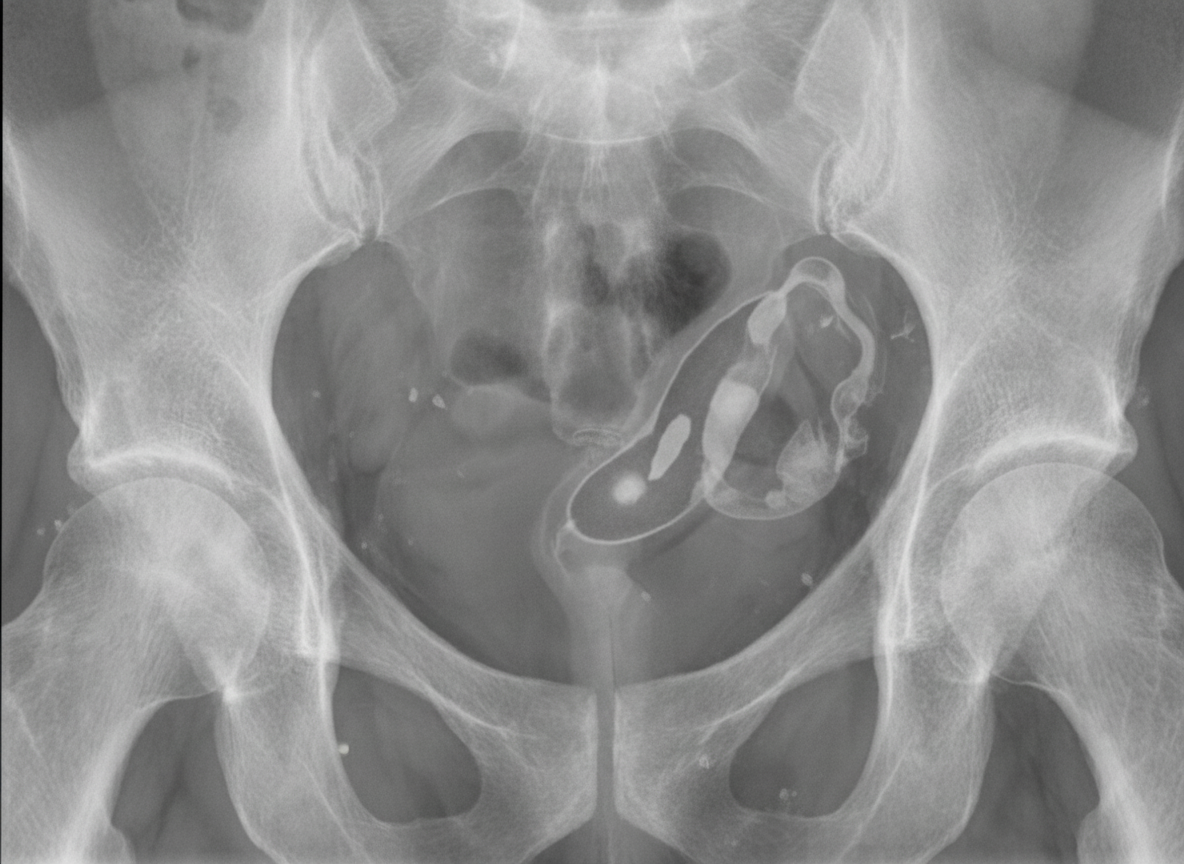
A hysterosalpingogram shows which of the following findings suggestive of a potential cause of infertility?
All are side effects of clomiphene citrate except?
Chlamydia infection can cause infertility due to which of the following complications?
A 25-year-old male underwent semen analysis. Results show: sperm count - 15 million/ml; pH - 7.5; volume - 2 ml; no agglutination is seen. Morphology shows 60% normal and 60% motile sperms. What is the most likely diagnosis?
Which hormone best indicates ovarian reserve?
Fallopian tube dysmotility is seen in which of the following conditions?
What is the best investigation to assess tubal patency?
Practice by Chapter
Reproductive Physiology
Practice Questions
Evaluation of the Infertile Couple
Practice Questions
Male Factor Infertility
Practice Questions
Female Factor Infertility
Practice Questions
Ovulatory Disorders
Practice Questions
Tubal and Peritoneal Factors
Practice Questions
Uterine Factors
Practice Questions
Unexplained Infertility
Practice Questions
Assisted Reproductive Technologies
Practice Questions
Psychological Aspects of Infertility
Practice Questions
Want unlimited practice?
Get full access to all questions, explanations, and performance tracking.
Scan to download app