Most common site for fertilization?
In low ovarian reserve, anti-Müllerian hormone level will be:
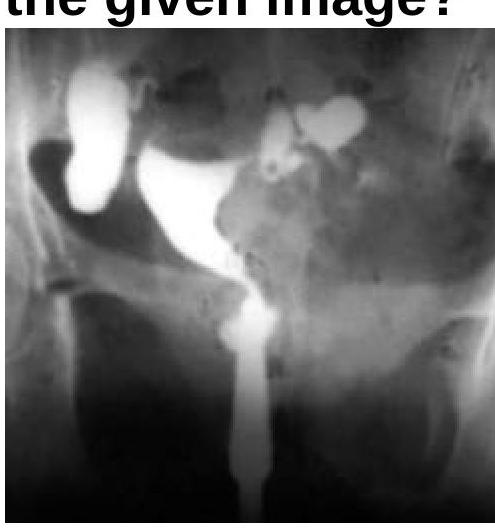
What will be the Hysterosalpingogram (HSG) finding?
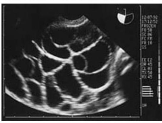
What is meant by Superfecundation?
The window of implantation occurs at which of the following time periods after fertilization?
Which of the following is not considered a marker of ovarian reserve?
What is the most appropriate management for a 28-year-old hemodynamically stable patient with mild abdominal pain and an unruptured tubal ectopic pregnancy measuring 2.5 x 3 cm, with β-hCG level of 8500 mIU/mL, visible fetal cardiac activity, and who desires future fertility?
On which day LH & FSH should be measured?
Ovarian reserve is best indicated by
What does teratozoospermia refer to?
Explanation: ***Ampulla*** - The **ampulla** of the **fallopian tube** is the widest and longest section, providing an ideal environment for the sperm and ovum to meet. - Fertilization most commonly occurs here, as it allows sufficient time for sperm capacitation and interaction with the egg. *Isthmus* - The **isthmus** is a narrow, thick-walled section of the fallopian tube, closer to the uterus. - While sperm may pass through here, it is not the primary site for fertilization. *Intramural* - The **intramural** (or interstitial) part is the segment of the fallopian tube that passes through the muscular wall of the uterus. - This narrowest part is not conducive to fertilization. *Fimbriae* - The **fimbriae** are finger-like projections at the end of the fallopian tube that capture the ovum after ovulation. - Their role is to direct the egg into the tube, not to be the site of fertilization.
Explanation: ***<1 ng/ml*** - **Low ovarian reserve** is characterized by a reduced quantity and quality of oocytes, which is reflected by **low anti-Müllerian hormone (AMH)** levels. - An AMH level of **<1 ng/ml** is generally indicative of a significantly diminished ovarian reserve. *1-4 ng/ml* - This range typically represents a **normal or adequate ovarian reserve**, suggesting a healthy number of remaining follicles. - Women with AMH levels in this range usually have a good response to ovarian stimulation in fertility treatments. *>7 ng/ml* - AMH levels **>7 ng/ml** are considered **high** and can be indicative of conditions such as **polycystic ovary syndrome (PCOS)**. - While high AMH suggests a large number of growing follicles, it is not associated with low ovarian reserve. *>10 ng/ml* - This level is also considered **very high**, even more suggestive of conditions like **PCOS** or a very robust ovarian reserve. - It is far from indicating a diminished or low ovarian reserve.
Explanation: ***Hydrosalpinx*** - The image, likely a hysterosalpingogram (HSG), shows a **dilated and fluid-filled fallopian tube** with no spillage of contrast into the peritoneal cavity, which is characteristic of hydrosalpinx. - The **contrast media fills the tubal lumen** but is unable to egress, indicating distal tubal obstruction and fluid accumulation. *Cornual block* - A cornual block would present as **obstruction at the uterine ostium** of the fallopian tube, preventing contrast from entering the tubal lumen. - In this image, contrast has clearly entered and dilated the fallopian tube, ruling out a cornual block. *Normal findings* - Normal HSG findings would show **patent fallopian tubes** with free spill of contrast into the peritoneal cavity. - The visible **dilation** and **lack of spill** in the image are distinctly abnormal. *Bicornuate uterus* - A bicornuate uterus is a **congenital uterine anomaly** characterized by two separate uterine horns. - While the uterus appears somewhat irregular, the dominant feature is the dilated fallopian tube, which is not a hallmark of a bicornuate uterus.
Explanation: ***Fertilization of two or more ova in different intercourses in same menstrual cycle*** - **Superfecundation** occurs when two or more ova released during the same menstrual cycle are fertilized by sperm from **separate acts of coitus**. - This can lead to **dizygotic twins** or multiples conceived at different times within a short window, potentially from different biological fathers. *Fertilization of two or more ova in one intercourse* - This scenario describes the fertilization of multiple ova within a **single sexual encounter**, often leading to **dizygotic multiples** but not superfecundation. - Superfecundation specifically implies fertilization from **separate instances of intercourse**. *Fertilization of ova and then its division* - This describes the formation of **monozygotic (identical) twins**, where a single fertilized ovum (zygote) later splits into two or more embryos. - It is distinct from superfecundation, which involves fertilization of **multiple ova**. *Fertilization of second ovum after first one is already implanted* - This describes **superfetation**, a rare phenomenon where a new pregnancy (fertilization and conception) occurs **while already pregnant** from a previous cycle. - Superfecundation, conversely, involves **multiple conceptions within the same menstrual cycle**, not across different cycles.
Explanation: ***6-10 days*** - The uterus is most receptive to implantation during the **"window of implantation,"** which occurs roughly **6 to 10 days post-fertilization**, coinciding with the mid-luteal phase. - During this period, the **endometrial lining** undergoes specific changes, guided by hormonal signals from **progesterone**, making it optimal for the blastocyst to attach. *12 days* - While implantation can still occur, the **peak receptivity window** is generally considered to be narrower, between 6 and 10 days. - By day 12, changes in the **endometrial environment** may start to reduce the likelihood of successful implantation. *12 weeks* - **12 weeks** refer to the end of the first trimester of pregnancy and is far too late for the initial implantation event. - Implantation must have occurred much earlier for a viable pregnancy at this stage. *6 weeks* - **6 weeks** refers to an established pregnancy, at which point implantation would have occurred several weeks prior. - The process of implantation is completed within the first two weeks post-fertilization.
Explanation: ***Inhibin A*** - **Inhibin A** levels primarily rise during the mid to late luteal phase and are involved in regulating FSH, but they are not a reliable or commonly used marker for **ovarian reserve**. - Its fluctuations are more indicative of the presence of a **corpus luteum** and short-term ovarian function rather than the total follicular pool. *Inhibin B* - **Inhibin B** is produced by granulosa cells of small antral follicles and is an important marker of **ovarian reserve**. - It inversely correlates with **FSH** levels in the early follicular phase, reflecting the number of developing follicles. *Ovarian volume* - **Ovarian volume**, particularly when measured by ultrasound, can be an indicator of **ovarian reserve**. - Smaller ovarian volume is generally associated with a reduced number of **antral follicles** and lower ovarian reserve. *Anti-Müllerian Hormone (AMH)* - **AMH** is a well-established and highly reliable marker of **ovarian reserve**, produced by the granulosa cells of preantral and small antral follicles. - Its levels correlate directly with the total number of remaining **primordial follicles** and are relatively stable throughout the menstrual cycle.
Explanation: ***Laparoscopic salpingostomy*** - This patient desires future fertility, making **salpingostomy** (tube-preserving surgery) the most appropriate management. - Salpingostomy involves making an incision in the fallopian tube, removing the ectopic pregnancy, and leaving the tube intact to preserve fertility potential. - While the presence of **fetal cardiac activity** and **β-hCG of 8500 mIU/mL** contraindicate medical management, they do not contraindicate conservative surgical management in a hemodynamically stable patient. - The patient meets criteria for conservative surgery: hemodynamically stable, unruptured ectopic, and desires future fertility. *Methotrexate therapy* - This patient has **absolute contraindications for methotrexate**: β-hCG level >5000 mIU/mL (here 8500) and presence of **fetal cardiac activity**. - Methotrexate is only suitable for hemodynamically stable patients with ectopic mass <3.5-4 cm, β-hCG <5000 mIU/mL, no fetal cardiac activity, and normal liver/renal function. - The high β-hCG and cardiac activity indicate a viable ectopic pregnancy that is unlikely to respond to medical management. *Laparoscopic salpingectomy* - Salpingectomy involves **complete removal of the affected fallopian tube**, which significantly reduces future fertility if this is the only functional tube or if the contralateral tube is damaged. - This option is preferred when: the tube is severely damaged, there is uncontrolled bleeding, recurrent ectopic in the same tube, or the patient does not desire future fertility. - Since this patient **specifically desires future fertility** and is hemodynamically stable with an unruptured ectopic, salpingostomy (tube preservation) is preferred over salpingectomy. *Expectant management* - Expectant management requires **very low or declining β-hCG levels** (typically <1000-1500 mIU/mL), absence of fetal cardiac activity, and very small ectopic size (<2 cm). - This patient has β-hCG of 8500 mIU/mL with **visible fetal cardiac activity**, indicating a viable growing ectopic pregnancy with high rupture risk. - These findings make expectant management unsafe and inappropriate.
Explanation: ***1-3rd day*** - Measuring **LH** (Luteinizing Hormone) and **FSH** (Follicle-Stimulating Hormone) on cycle days 1-3 provides a baseline assessment of **ovarian reserve** and pituitary function. - At this early follicular phase, hormone levels are relatively stable and reflect the intrinsic **gonadal feedback** mechanisms before significant follicular development begins. *7th day* - By day 7, **follicular development** is usually well underway, and FSH levels might be decreasing as a dominant follicle is selected. - Measuring hormones on this day would not provide an accurate baseline assessment, as the levels are already influenced by **follicular growth**. *14th day* - Day 14 is often associated with the **LH surge** that triggers ovulation, making it unsuitable for a baseline assessment of ovarian reserve. - FSH levels would also be significantly different from the early follicular phase due to the ongoing **ovarian cycle events**. *10th day* - On day 10, **estrogen levels** are typically rising, which would already be providing negative feedback to the pituitary, affecting FSH and LH levels. - This timing would not be ideal for assessing baseline hormone levels for **fertility evaluations**.
Explanation: ***Anti-Müllerian Hormone (AMH)*** - **AMH is currently considered the best single biochemical marker** for assessing ovarian reserve - Produced by **granulosa cells of preantral and small antral follicles**, directly reflecting the size of the primordial follicle pool - **Cycle-independent** - can be measured at any time during the menstrual cycle - **More sensitive and specific** than FSH for detecting diminished ovarian reserve - **Minimal inter-cycle and intra-cycle variability**, providing consistent and reliable results - Widely used in **fertility assessment, IVF protocols**, and predicting ovarian response to stimulation *Follicle-stimulating hormone (FSH)* - Elevated **early follicular phase FSH** (measured on day 3) indicates diminished ovarian reserve - Historically the most commonly used marker, but **less sensitive than AMH** - **Cycle-dependent** - must be measured on specific days (day 2-4 of cycle) - A **late marker** - rises only when ovarian reserve is already significantly diminished - Still clinically useful and widely available, but not the "best" indicator *Luteinizing hormone (LH)* - **LH** primarily triggers ovulation and does not directly reflect ovarian reserve - Elevated in conditions like **PCOS** but does not assess the quantity or quality of remaining follicles - Not a reliable indicator of overall ovarian reserve *LH/FSH ratio* - An elevated **LH/FSH ratio** (>2:1 or >3:1) is associated with **Polycystic Ovary Syndrome (PCOS)** - Reflects anovulation and hormonal imbalance, not the number or viability of ovarian follicles - Does not assess ovarian reserve capacity
Explanation: ***Morphologically defective sperm*** - **Teratozoospermia** specifically refers to the presence of an unusually high percentage of **abnormally shaped sperm** in an ejaculate. - These malformations can affect the **head, midpiece, or tail** of the sperm, potentially impairing its ability to fertilize an egg. *Low sperm count* - This condition is known as **oligozoospermia**, which refers to a sperm concentration below the normal range. - While low sperm count can affect fertility, it is distinct from issues with sperm morphology. *Sperm with abnormal motility* - This condition is called **asthenozoospermia**, characterized by reduced or absent sperm movement. - Poor motility impacts the sperm's ability to reach and penetrate the egg, but it is not directly related to sperm shape. *Absence of sperm in semen* - The complete absence of sperm in the ejaculate is known as **azoospermia**. - This is a severe form of male infertility, different from having sperm with structural defects.
Reproductive Physiology
Practice Questions
Evaluation of the Infertile Couple
Practice Questions
Male Factor Infertility
Practice Questions
Female Factor Infertility
Practice Questions
Ovulatory Disorders
Practice Questions
Tubal and Peritoneal Factors
Practice Questions
Uterine Factors
Practice Questions
Unexplained Infertility
Practice Questions
Assisted Reproductive Technologies
Practice Questions
Psychological Aspects of Infertility
Practice Questions
Get full access to all questions, explanations, and performance tracking.
Start For Free