Most common cause of early abortion?
Least common site of ectopic pregnancy in the fallopian tubes is?
In which phase of the menstrual cycle should a tubal patency test be performed?
Fertilized ovum reaches the uterus at what day of menstrual cycle?
Fertilization usually occurs in which part of fallopian tube?
Within what timeframe after ovulation does fertilization typically occur?
A female presents with 6 weeks of amenorrhea, experiencing vaginal bleeding and slight abdominal pain. A urine pregnancy test is positive, and her hCG level is 2800 IU/L. A mass measuring 3 x 2.5 cm is observed on the left adnexa. She is hemodynamically stable. What is the most appropriate management for this patient?
A 23-year-old woman accompanied by her mother-in-law comes to the infertility clinic. She has been having regular intercourse for 6 months but is not able to conceive. What is the next best step?
A lady on treatment for infertility developed ascites, abdominal pain, and dyspnea. The ultrasound image is shown below. What is the most likely diagnosis?

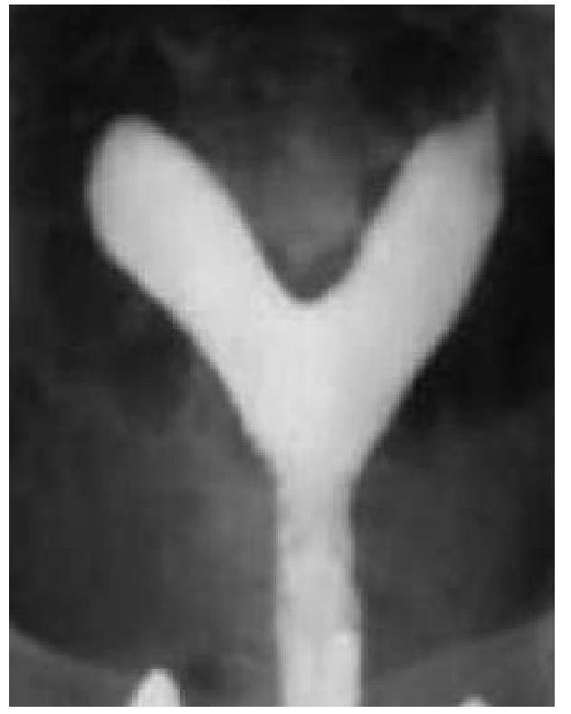
A 20 year old woman is evaluated for primary infertility. Hysterosalpingography was done and reveals an anomaly. What is the anomaly seen in the image?

Explanation: ***Correct: Genetic*** - **Chromosomal abnormalities**, such as aneuploidy, are the most frequent cause of **spontaneous abortions**, especially early in pregnancy (accounting for **50-60% of first-trimester losses**). - These genetic errors often lead to **non-viable embryos**, resulting in early pregnancy loss. - Most common abnormalities include **trisomies** (especially trisomy 16), **monosomy X**, and **triploidy**. *Incorrect: Maternal* - While maternal factors like **diabetes** or **thyroid dysfunction** can contribute to abortion, they are less common causes of early abortion compared to genetic issues. - Systemic maternal health problems usually account for a smaller percentage of all miscarriages. *Incorrect: Immunologic* - **Autoimmune disorders** like **antiphospholipid syndrome** can cause recurrent pregnancy loss, but they are not the primary cause of the majority of early, sporadic abortions. - These are typically considered in cases of **recurrent miscarriages**, not usually the first or early spontaneous abortion. *Incorrect: Anatomic abnormalities* - **Uterine anomalies** (e.g., septate uterus) or **cervical incompetence** can lead to recurrent pregnancy loss, particularly in the later first or second trimester. - However, they are less frequently the cause of very early, isolated miscarriages compared to genetic factors.
Explanation: ***Interstitium*** - The **interstitial portion** of the fallopian tube is the narrowest and thickest part, making it the least common site for an ectopic pregnancy within the tube itself. - While less frequent, interstitial pregnancies are associated with a **higher risk of rupture** and hemorrhage due to the surrounding myometrial tissue. *Ampulla* - The **ampulla** is the most common site of ectopic pregnancy in the fallopian tube, accounting for about 80% of all tubal ectopics. - Its wider diameter and convoluted lumen easily trap the fertilized egg. *Infundibulum* - The **infundibulum**, or fimbrial end, is a relatively common site for ectopic pregnancies after the ampulla, accounting for about 5% of cases. - Pregnancies here are often referred to as **fimbrial ectopic pregnancies**. *Isthmus* - The **isthmus** is the second most common site for tubal ectopic pregnancies, accounting for approximately 12% of cases. - This section is narrower than the ampulla but still more hospitable than the interstitial portion.
Explanation: ***Preovulatory*** - Performing a **tubal patency test** during the **preovulatory phase** (also known as the late follicular phase) minimizes the risk of interfering with a potential early pregnancy, as ovulation has not yet occurred. - The endometrium is thinner during this time, potentially allowing for better visualization and reducing the chance of discomfort. *Menstrual* - Performing the test during the **menstrual phase** is typically avoided due to the presence of blood and tissue, which can obscure visualization and increase the risk of infection. *Premenstrual* - The **premenstrual phase** occurs after ovulation and before menstruation, making it possible that a pregnancy could be inadvertently disrupted if conception has occurred. - The **endometrium is thick** during this phase, which could also interfere with visualization and test interpretation. *Luteal* - The **luteal phase** begins after ovulation, meaning there is a higher risk of disrupting an early, undetected **pregnancy** if the test is performed during this time. - Similar to the premenstrual phase, the **endometrium is secretory and thick**, which can complicate the procedure and interpretation.
Explanation: ***20th*** - **Fertilization** occurs around day 14 of a 28-day cycle in the ampulla of the fallopian tube. - The fertilized ovum (zygote) takes approximately **3-4 days** to travel through the fallopian tube, reaching the uterine cavity around **day 17-18**. - By day 20-21, the blastocyst begins **implantation** into the endometrium. - Note: Some references cite day 20 as it marks the transition period when the blastocyst is fully in the uterus and beginning implantation. *6th* - This is far too early, as ovulation typically hasn't occurred yet by day 6. - On day 6, a woman is in the **follicular phase** with developing ovarian follicles. - Fertilization cannot occur before ovulation (around day 14). *14th* - Day 14 is typically the day of **ovulation** and fertilization in a 28-day cycle. - At this point, the egg has just been released from the ovary and is in the **fallopian tube**. - The fertilized ovum requires 3-4 days of transport to reach the uterine cavity. *25th* - By day 25, implantation would have already been completed (implantation occurs around day 20-21). - The embryo would be producing **hCG** and establishing placental connections. - If no pregnancy occurred, the corpus luteum would be regressing and the endometrium preparing for **menstruation**.
Explanation: ***Ampulla*** - The **ampulla** is the widest and longest part of the fallopian tube, making it the most common site for **fertilization** to occur. - Sperm typically meet the oocyte here after ovulation and transport through the fimbriae. *Fimbrial end* - The **fimbriae** are finger-like projections at the end of the fallopian tube that **capture the ovulated oocyte**. - While essential for collecting the egg, it is not the primary site where fertilization takes place. *Interstitium* - The **interstitium** (or intramural part) is the segment of the fallopian tube that passes through the **uterine wall**. - It is too narrow and close to the uterus for fertilization to normally occur there. *Isthmus* - The **isthmus** is the narrow, muscular part of the fallopian tube connecting the ampulla to the uterus. - While sperm may reside here for a short period before fertilization, it is not the typical site where the sperm and egg unite.
Explanation: ***12-24 hours*** - The **ovum** is viable for fertilization for a relatively short period, typically **12 to 24 hours** after its release during ovulation. - For fertilization to occur, **sperm** must be present in the fallopian tube within this critical window. *5-6 days* - This timeframe is more characteristic of the **duration that sperm can survive** in the female reproductive tract, rather than the viability of the ovum for fertilization. - If sperm are present in the reproductive tract up to **5-6 days before ovulation**, they may still fertilize the egg once it's released. *8-12 days* - This period extends significantly beyond the **viability of the ovum** for fertilization. - An ovum released during ovulation will have degenerated and will no longer be capable of being fertilized after 24 hours. *> 12 days* - Fertilization cannot occur this late after ovulation as the **egg would have long since degenerated**. - This timeframe is not relevant to the process of fertilization itself.
Explanation: ***Single-dose methotrexate injection*** - The patient presents with a **hemodynamically stable ectopic pregnancy**, as suggested by the positive pregnancy test, amenorrhea, vaginal bleeding, abdominal pain, an hCG level of 2800 IU/L, and an adnexal mass. - A single-dose methotrexate injection is the **first-line medical management** for ectopic pregnancies in stable patients, particularly when hCG levels are typically below 5000 IU/L and the mass size is less than 4 cm (or 3.5 cm in some guidelines). *Oral methotrexate* - **Oral methotrexate** is not typically used for the treatment of ectopic pregnancy due to unpredictable absorption and less reliable therapeutic efficacy compared to intramuscular administration. - The **intramuscular route** is preferred to ensure consistent systemic exposure and effectiveness in dissolving the ectopic gestation. *Serial methotrexate + leucovorin rescue* - **Leucovorin rescue** is used to mitigate the adverse effects of high-dose methotrexate, typically in cancer chemotherapy, and is not indicated for the standard treatment of ectopic pregnancy. - Serial doses of methotrexate without leucovorin may be given if the initial single dose fails, but **leucovorin is not part of the standard initial treatment protocol** for ectopic pregnancy. *Salpingectomy* - **Salpingectomy (surgical removal of the fallopian tube)** is indicated for ectopic pregnancies that are **hemodynamically unstable**, ruptured, too large for medical management (e.g., >4 cm), or in cases where medical management with methotrexate fails or is contraindicated. - Since this patient is **hemodynamically stable** and meets the criteria for medical management, surgery is not the most appropriate initial management.
Explanation: ***Reassure and review the couple after 6 months*** - Infertility is defined as the inability to conceive after **12 months** of regular, unprotected intercourse in women under 35 years old. For women aged 35 or older, this period is 6 months. - Since the patient is 23 years old and has been trying for only 6 months, she does not yet meet the diagnostic criteria for infertility. The appropriate action is to advise them to continue trying and to return for evaluation if conception does not occur after a full year. *Semen analysis for husband* - While a semen analysis is a crucial initial step in an infertility workup, it is premature at this stage given the duration of attempted conception. - It would be appropriate to order this test after the couple has met the criteria for infertility (12 months for women under 35). *Hysterolaparoscopy* - This is an invasive procedure typically reserved for more advanced stages of an infertility workup, especially when suspected pathologies like endometriosis or tubal factor infertility are present. - It is not indicated as an initial step for a couple who has only been trying to conceive for 6 months and does not yet meet the definition of infertility. *Diagnostic hysteroscopy* - A diagnostic hysteroscopy is used to visualize the inside of the uterus to identify intrauterine pathologies that could contribute to infertility. - Like hysterolaparoscopy, it is an invasive diagnostic tool and should only be considered after initial, less invasive investigations have been performed and the couple meets the criteria for infertility.
Explanation: ***OHSS*** - The clinical presentation of a woman undergoing infertility treatment who develops **ascites**, **abdominal pain**, and **dyspnea** is classic for **Ovarian Hyperstimulation Syndrome (OHSS)**. The ultrasound image showing massively enlarged, multicystic ovaries with numerous follicles further confirms this diagnosis. - OHSS is an iatrogenic complication of **ovarian stimulation**, where ovaries become hyperstimulated, leading to **capillary permeability** and fluid shifts, resulting in ascites and potentially pleural effusions causing dyspnea. *PCOS* - While Polycystic Ovarian Syndrome (PCOS) involves multiple small follicles (usually 12 or more per ovary, each 2-9 mm in diameter), it typically does not present with acute symptoms like **ascites** and **dyspnea** unless severe OHSS occurs after ovulation induction in a woman with PCOS. - The ovaries in PCOS are generally smaller or normal size compared to the massively enlarged ovaries seen in the image, and the presence of significant ascites and dyspnea is not a direct feature of PCOS itself. *Theca lutein cyst* - **Theca lutein cysts** are usually **bilateral**, **multiloculated ovarian cysts** that result from exaggerated stimulation by **human chorionic gonadotropin (hCG)**, often seen in conditions like **gestational trophoblastic disease** or **multiple gestations**. - While they can be large and multicystic, the presentation with acute ascites and dyspnea in the context of infertility treatment points more specifically to OHSS. *Mucinous cystadenomas* - **Mucinous cystadenomas** are benign **epithelial ovarian tumors** that can grow very large and be multiloculated, but they are not typically associated with infertility treatment or the acute systemic symptoms of **ascites** and **dyspnea** via capillary leak syndrome. - Their presence would be coincidental rather than a direct complication of infertility therapy, and their fluid is usually thick and mucinous, enclosed within the cyst wall rather than causing diffuse fluid extravasation.
Explanation: ***Septate uterus*** - The image exhibits a **single uterine cavity** with a **septum** or indentation extending downwards, splitting the cavity into two distinct portions superiorly. - This configuration, particularly with an external contour that is typically **convex or flat**, is characteristic of a septate uterus, which is often associated with recurrent pregnancy loss and infertility. *Uterine didelphys* - This anomaly involves **two completely separate uteri**, each with its own cervix and often a separate vagina. - The image clearly shows a single main uterine body that then divides superiorly, not two entirely distinct uteri. *Bicornuate uterus* - A bicornuate uterus typically has two uterine horns that are **divergent externally**, creating a **deep indentation** on the external contour of the fundus. - While it also involves a divided uterine cavity, the external contour in the image appears more convex or flat, which is less consistent with a bicornuate uterus where the outer fundal contour is notably indented. *Unicornuate uterus* - This anomaly results from the **failure of one Müllerian duct to develop**, leading to a uterus that has only one horn and one fallopian tube. - The image presents a uterus with two distinct horns, ruling out a unicornuate uterus.
Reproductive Physiology
Practice Questions
Evaluation of the Infertile Couple
Practice Questions
Male Factor Infertility
Practice Questions
Female Factor Infertility
Practice Questions
Ovulatory Disorders
Practice Questions
Tubal and Peritoneal Factors
Practice Questions
Uterine Factors
Practice Questions
Unexplained Infertility
Practice Questions
Assisted Reproductive Technologies
Practice Questions
Psychological Aspects of Infertility
Practice Questions
Get full access to all questions, explanations, and performance tracking.
Start For Free