
Fertility and Infertility — MCQs

On this page
Which of the following is not a risk factor for ectopic pregnancy?
Which of the following is NOT an indication for hysterosalpingography?
Clomiphene citrate is primarily used for treating which condition in women?
In which phase of the menstrual cycle should a tubal patency test be performed?
A 23-year-old woman accompanied by her mother-in-law comes to the infertility clinic. She has been having regular intercourse for 6 months but is not able to conceive. What is the next best step?
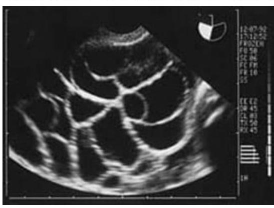
A lady on treatment for infertility developed ascites, abdominal pain, and dyspnea. The ultrasound image is shown below. What is the most likely diagnosis?
In low ovarian reserve, anti-Müllerian hormone level will be:
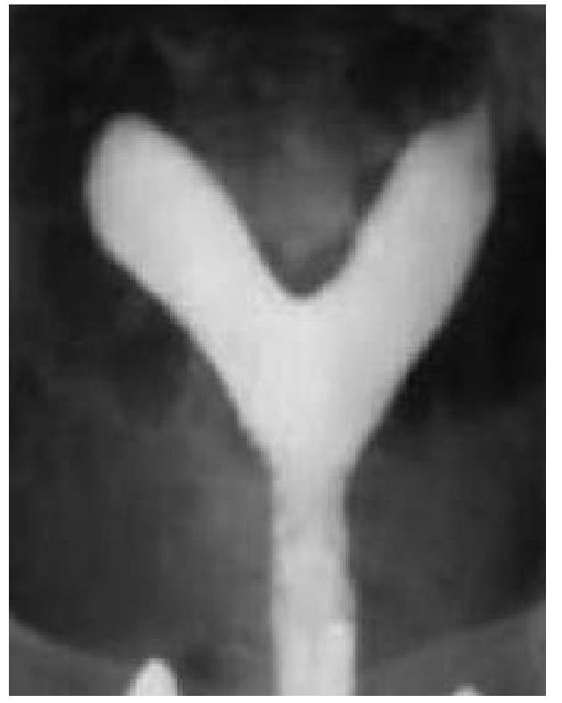
A 20 year old woman is evaluated for primary infertility. Hysterosalpingography was done and reveals an anomaly. What is the anomaly seen in the image?
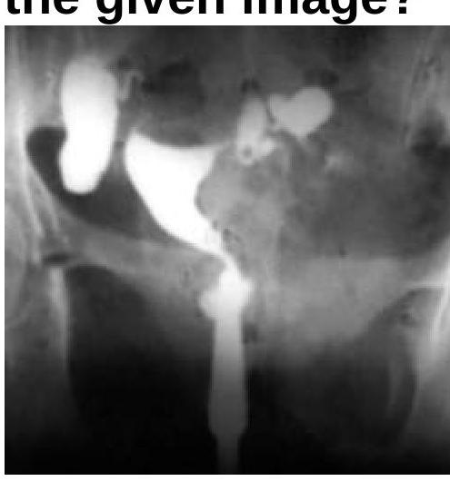
What will be the Hysterosalpingogram (HSG) finding?
The window of implantation occurs at which of the following time periods after fertilization?
Practice by Chapter
Reproductive Physiology
Practice Questions
Evaluation of the Infertile Couple
Practice Questions
Male Factor Infertility
Practice Questions
Female Factor Infertility
Practice Questions
Ovulatory Disorders
Practice Questions
Tubal and Peritoneal Factors
Practice Questions
Uterine Factors
Practice Questions
Unexplained Infertility
Practice Questions
Assisted Reproductive Technologies
Practice Questions
Psychological Aspects of Infertility
Practice Questions
Want unlimited practice?
Get full access to all questions, explanations, and performance tracking.
Scan to download app