
Fertility and Infertility — MCQs

On this page
A married patient for 3 years is unable to conceive despite having regular menstrual cycles. Her husband's semen analysis and hormonal profile are normal. What is the optimal timing in the menstrual cycle to perform an endometrial biopsy for infertility evaluation?
Which of the following is NOT an essential criterion according to WHO for normal semen analysis?
In which case homologous artificial insemination is used in females?
Decreased motility of the fallopian tube is seen in which condition?
Preimplantation genetic diagnosis is used for which of the following?
Infertility issues associated with leiomyoma can be addressed by which of the following?
A 35-year-old female patient presents to the clinic for evaluation of her fertility status. She has been trying to conceive for over a year without success. Which of the following is the single best test for assessing her ovarian reserve?
Ovarian drilling is done in which of the following conditions?
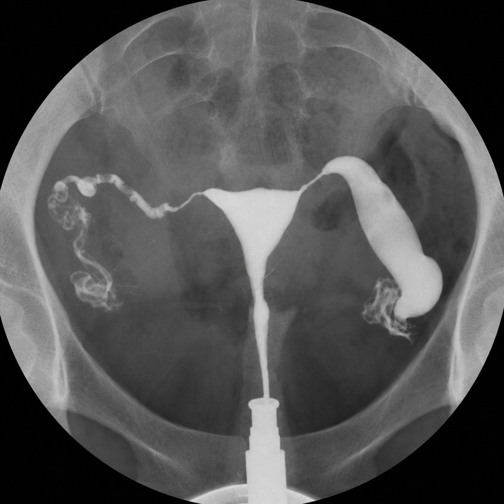
Which of the following is seen in the HSG given below?
Which of the following statements about hysterosalpingography, as a diagnostic procedure, are correct? I. Tubal patency assessment following tuboplasty operation II. Diagnosis of uterine synechiae III. Detection of IUD IV. Diagnosis of subserosal fibroid Select the answer using the code given below :
Practice by Chapter
Reproductive Physiology
Practice Questions
Evaluation of the Infertile Couple
Practice Questions
Male Factor Infertility
Practice Questions
Female Factor Infertility
Practice Questions
Ovulatory Disorders
Practice Questions
Tubal and Peritoneal Factors
Practice Questions
Uterine Factors
Practice Questions
Unexplained Infertility
Practice Questions
Assisted Reproductive Technologies
Practice Questions
Psychological Aspects of Infertility
Practice Questions
Want unlimited practice?
Get full access to all questions, explanations, and performance tracking.
Scan to download app