Hysteroscopic excision is indicated for which of the following conditions?
Endosalpingitis is best diagnosed by?
35 yr old with 4 months amenorrhea with increased FSH, decreased estrogen. What is the diagnosis?
Chlamydia causes:
A 30-year-old woman presents with primary infertility for 2 years. Her menstrual cycles are irregular (35-45 days). Investigations reveal FSH 15 IU/L, AMH 0.5 ng/mL. Semen analysis is normal and HSG shows patent tubes. What is the most appropriate first-line treatment?
What is the most reliable test to confirm ovulation after it has occurred?
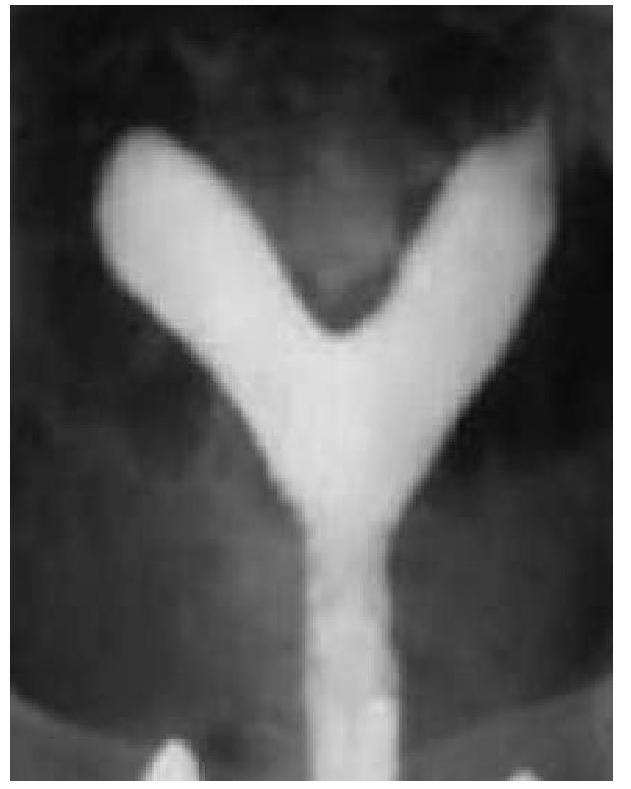
A 20 year old woman is evaluated for primary infertility. Hysterosalpingography was done and reveals an anomaly. What is the anomaly seen in the image?

Which of the following is a cause of male infertility?
Lady presents with infertility and diagnosed with bilateral cornual block on hysterosalpingography. What is the next step?
In a couple, which of the following investigations are included in the initial work-up for infertility?
Explanation: ***Submucous fibroid*** - **Hysteroscopic myomectomy** is the **gold standard treatment** for submucous fibroids that protrude into the uterine cavity. - Type 0 and Type I submucous fibroids are **ideal candidates** for hysteroscopic excision, as they are directly accessible through the cervix. - This **minimally invasive approach** preserves the uterus and fertility while effectively removing the fibroid. - Submucous fibroids commonly cause **heavy menstrual bleeding** and require definitive excision for symptom relief. *Subserous fibroid* - Located on the **outer surface of the uterus**, projecting into the peritoneal cavity. - Not accessible via hysteroscopy; requires **laparoscopy or laparotomy** for removal. - The hysteroscope cannot reach these fibroids as they are outside the uterine cavity. *Uterine fundus fibroid* - These are typically **intramural fibroids** embedded within the uterine muscle wall. - Not suitable for hysteroscopic excision unless they have a significant submucous component. - Would require **laparoscopic or open myomectomy** depending on size and location. *Endometrial polyp* - **Hysteroscopic polypectomy** is indeed the standard treatment for endometrial polyps. - While this is a valid indication for hysteroscopy, endometrial polyps are typically **smaller, benign lesions** that are easier to remove. - In the context of this question, **submucous fibroid** is the more specific answer as it represents a more complex pathology where hysteroscopic excision is both technically demanding and clinically significant. - Both are correct indications, but submucous fibroid is the **primary surgical indication** that best demonstrates the therapeutic value of hysteroscopic excision for larger structural abnormalities.
Explanation: ***Laparoscopy*** - **Laparoscopy** is the **gold standard** for diagnosing endosalpingitis as it allows direct visualization of the fallopian tubes, pelvic organs, and peritoneal cavity. - It enables identification of **inflammation, adhesions, tubal edema, and purulent exudate** characteristic of endosalpingitis. - It also permits **tissue sampling** for histopathological confirmation and culture of infectious agents. - Laparoscopy has high sensitivity and specificity for diagnosing pelvic inflammatory disease (PID) and its complications. *X-Ray abdomen* - An **X-ray abdomen** provides limited information regarding soft tissue structures like the fallopian tubes. - It is primarily used for visualizing bones or detecting gross abnormalities like bowel obstruction or free air. - It **cannot directly diagnose endosalpingitis** or provide detailed images of adnexal structures. *Hysterosalpingography* - **Hysterosalpingography (HSG)** is an imaging technique used to assess the patency and contour of the fallopian tubes and uterine cavity by injecting contrast dye. - While it can detect **tubal occlusion or hydrosalpinx**, it cannot visualize external tubal inflammation, adhesions, or the peritoneal surface. - HSG is more useful for evaluating **tubal patency in infertility workup** rather than diagnosing acute inflammation. *Hystero-laparoscopy* - This term refers to **combined hysteroscopy and laparoscopy** performed together. - While the laparoscopic component can diagnose endosalpingitis, **hysteroscopy** (visualization of the uterine cavity) adds no additional value for diagnosing tubal inflammation. - For endosalpingitis specifically, **laparoscopy alone** is sufficient and is the most direct diagnostic approach.
Explanation: ***Premature ovarian failure*** - The combination of **amenorrhea** for 4 months in a 35-year-old, with **increased FSH** and **decreased estrogen**, is characteristic of premature ovarian failure, indicating the ovaries are no longer responding to FSH stimulation. - This condition signifies the cessation of ovarian function before the age of 40, leading to menopausal symptoms and infertility. *Pituitary dysfunction* - Pituitary dysfunction might lead to **decreased FSH** (hypogonadotropic hypogonadism) due to insufficient stimulation of the ovaries, not increased FSH. - In cases of pituitary adenomas, increased prolactin can cause amenorrhea, but FSH would not be elevated in the manner described. *Hypothalamic dysfunction* - Hypothalamic dysfunction, such as **functional hypothalamic amenorrhea**, typically presents with **low or normal FSH and LH levels** (hypogonadotropic hypogonadism) due to reduced GnRH pulsatility. - This condition is often associated with stress, excessive exercise, or low body weight, and would not cause elevated FSH as seen here. *Polycystic Ovary Syndrome* - **Polycystic Ovary Syndrome (PCOS)** is characterized by **anovulation**, resulting in amenorrhea or oligomenorrhea, but typically involves **elevated androgens** and a **high LH-to-FSH ratio**, with FSH levels generally normal or low, and estrogen levels often normal or slightly elevated. - It would not present with simultaneously high FSH and low estrogen, which points to ovarian failure rather than anovulation with intact ovarian reserve.
Explanation: **Trachoma and Lymphogranuloma venereum** - This is the **most comprehensive correct answer** as it lists two major diseases caused by different serovars of *Chlamydia trachomatis* - **Trachoma** (serovars A, B, Ba, C) - chronic keratoconjunctivitis and the leading infectious cause of blindness worldwide - **Lymphogranuloma venereum (LGV)** (serovars L1, L2, L3) - systemic sexually transmitted infection with inguinal lymphadenopathy - This answer captures both the **ocular** and **genital** manifestations of *C. trachomatis*, making it the best option *Trachoma* - While medically accurate, this is **incomplete** as it only mentions one disease - *C. trachomatis* causes multiple clinically significant diseases beyond trachoma - This option ignores LGV and other important chlamydial syndromes *Trachoma and Conjunctivitis* - This option is **redundant and misleading** - trachoma IS a specific type of chronic bacterial conjunctivitis - Listing both together suggests they are separate entities when trachoma is actually a subset of conjunctivitis - Misses other major non-ocular diseases like LGV, making it inferior to the correct answer *Conjunctivitis* - While *C. trachomatis* does cause conjunctivitis (trachoma, inclusion conjunctivitis), this is **too broad and non-specific** - Conjunctivitis has numerous causes (viral, bacterial, allergic) and this answer lacks specificity - Completely omits systemic/genital manifestations like LGV, urethritis, and PID
Explanation: ***Letrozole*** - This patient has **diminished ovarian reserve (DOR)** evidenced by **FSH 15 IU/L** (borderline elevated) and **AMH 0.5 ng/mL** (significantly low), along with **irregular cycles suggesting anovulation**. - **Letrozole**, an aromatase inhibitor, is the **preferred first-line ovulation induction agent** in patients with DOR who are anovulatory. It works by reducing estrogen production, leading to increased FSH release and follicular development. - **Advantages over clomiphene:** Letrozole has fewer anti-estrogenic effects on the endometrium and cervical mucus, making it superior in DOR patients where endometrial receptivity is crucial. - Given her young age (30 years) and evidence of some ovarian reserve (AMH 0.5, not undetectable), a trial of **2-3 cycles of ovulation induction** before proceeding to IVF is reasonable and cost-effective. *IVF with donor eggs* - This is **premature as first-line therapy**. While the patient has DOR, she still has detectable AMH (0.5 ng/mL), indicating some ovarian function remains. - The **first step** would be attempting conception with her own eggs through ovulation induction, and if that fails, **IVF with autologous (own) eggs** should be tried before considering donor eggs. - Donor egg IVF is typically reserved for patients with **premature ovarian failure**, very advanced age, or after repeated failed IVF cycles with own eggs. *Clomiphene citrate* - While clomiphene is an effective **ovulation induction agent**, it has significant **anti-estrogenic effects** on the endometrium (causing thinning) and cervical mucus (reducing quality). - In patients with **DOR**, where pregnancy rates are already compromised, these anti-estrogenic effects can further reduce success rates. - **Letrozole has been shown to have better pregnancy outcomes** in various populations, including those with DOR, making it the preferred first-line agent. *Gonadotropins* - Injectable gonadotropins (FSH/LH) directly stimulate follicular development and are more potent than oral agents. - However, they carry **higher risks**: multiple gestations (20-30%), ovarian hyperstimulation syndrome (OHSS), and significantly higher cost. - They are typically reserved as **second-line therapy** after failed response to oral ovulation induction agents, or as part of IVF protocols. - Starting with less aggressive, safer oral agents like letrozole is the **standard stepwise approach** in infertility management.
Explanation: ***Serum progesterone*** - A **serum progesterone level** of greater than **3 ng/mL (or 10 nmol/L)** in the mid-luteal phase (approximately 7 days after the presumed ovulation) reliably indicates that ovulation has occurred. - After ovulation, the **corpus luteum** forms and produces progesterone, causing a characteristic rise in its serum level. *Serum estrogen* - Estrogen levels **peak before ovulation** to trigger the LH surge and also rise during the luteal phase, but a single measurement is not a reliable indicator that ovulation has successfully occurred. - Estrogen levels can fluctuate due to various factors and do not directly confirm the **formation and function of a corpus luteum** as progesterone does. *Both serum estrogen and progesterone* - While both hormones are involved in the menstrual cycle, relying on both simultaneously for confirming *occurred* ovulation is not the most precise method. - A significant rise in **progesterone** *after* the presumed ovulatory event is the key reliable biomarker. *None of the options* - This option is incorrect because **serum progesterone** is a well-established and reliable test for confirming ovulation.
Explanation: ***Septate uterus*** - The image exhibits a **single uterine cavity** with a **septum** or indentation extending downwards, splitting the cavity into two distinct portions superiorly. - This configuration, particularly with an external contour that is typically **convex or flat**, is characteristic of a septate uterus, which is often associated with recurrent pregnancy loss and infertility. *Uterine didelphys* - This anomaly involves **two completely separate uteri**, each with its own cervix and often a separate vagina. - The image clearly shows a single main uterine body that then divides superiorly, not two entirely distinct uteri. *Bicornuate uterus* - A bicornuate uterus typically has two uterine horns that are **divergent externally**, creating a **deep indentation** on the external contour of the fundus. - While it also involves a divided uterine cavity, the external contour in the image appears more convex or flat, which is less consistent with a bicornuate uterus where the outer fundal contour is notably indented. *Unicornuate uterus* - This anomaly results from the **failure of one Müllerian duct to develop**, leading to a uterus that has only one horn and one fallopian tube. - The image presents a uterus with two distinct horns, ruling out a unicornuate uterus.
Explanation: ***All of the options*** - **Idiopathic**, **varicocele**, and **Yq11 microdeletion** are all recognized causes of male infertility, making this the most comprehensive and correct answer. - Male infertility can stem from a variety of factors, including genetic, structural, hormonal, and unexplained (idiopathic) causes. *Idiopathic* - Refers to cases where no specific cause for infertility can be identified despite thorough investigation, accounting for a significant proportion of male infertility. - This diagnosis is made by **exclusion** after ruling out other known causes. *Varicocele* - A common and treatable cause of male infertility, characterized by **dilated veins in the pampiniform plexus** of the scrotum. - Varicoceles can impair sperm production and function due to **increased scrotal temperature** and oxidative stress. *Yq11 microdeletion* - Refers to deletions in the **azoospermia factor (AZF) region** on the long arm of the Y chromosome, which are genetic causes of severe spermatogenic failure. - These deletions disrupt genes essential for sperm production, leading to conditions ranging from **oligozoospermia** (low sperm count) to **azoospermia** (absence of sperm).
Explanation: ***Laparoscopy and hysteroscopy*** - A **laparoscopy** allows for direct visualization of the fallopian tubes to confirm the tubal obstruction and assess for other pelvic pathology like **endometriosis** or **adhesions**. - A **hysteroscopy** can be performed concurrently to inspect the uterine cavity and the tubal ostia for any intracavitary abnormalities or to attempt **canalization of the cornual block**. *Tuboplasty* - **Tuboplasty** is a corrective surgical procedure for tubal obstruction, but it is typically considered *after* a definitive diagnosis and assessment of the block's extent have been made via diagnostic procedures. - Its success rate varies depending on the location and nature of the block, and it is not the immediate next step for diagnosis. *USG* - **Transvaginal ultrasonography (USG)** is a useful tool for evaluating uterine and ovarian morphology but is generally *not definitive* for diagnosing tubal patency or specific locations of tubal blockage. - While it can identify some pathologies, it cannot visualize the fallopian tubes with sufficient clarity to determine cornual obstruction. *IVF* - **In vitro fertilization (IVF)** is an *assisted reproductive technology* used to bypass tubal factor infertility, but it is a treatment option, not a diagnostic step. - It would be considered *after* a full diagnostic workup has confirmed the tubal blockage and other fertility factors, and after counseling regarding prognosis and success rates.
Explanation: ***Semen analysis, Tubal patency test, Ovulation test*** - This option correctly identifies the **key initial investigations** for both male and female factors in infertility: **semen analysis** for male fertility, **tubal patency test** for assessing fallopian tube function, and **ovulation test** to confirm female ovulatory cycles. - These tests are fundamental in a comprehensive initial infertility work-up as they address the most common causes of infertility. *Testicular biopsy, USG, Sperm penetration test* - While **testicular biopsy** and **sperm penetration test** are relevant, they are typically **second-line investigations** performed if initial tests (like semen analysis) are abnormal. - **Ultrasound (USG)** is a general imaging modality and not a primary, specific infertility test for both partners as listed. *Ovulation, tubal patency, Mantoux test* - **Ovulation** and **tubal patency** are essential, but the **Mantoux test** (for tuberculosis) is generally not part of the *initial routine* infertility work-up unless there is clinical suspicion or prevalence in the region. - The Mantoux test is specific for a particular infection, and not a broad screening test for infertility. *Semen analysis, CXR, Mantoux* - **Semen analysis** is appropriate, but a **Chest X-ray (CXR)** and **Mantoux test** are not routine initial investigations for infertility. - These tests would only be indicated if there were specific clinical signs or a history suggestive of underlying pulmonary or infectious disease.
Get full access to all questions, explanations, and performance tracking.
Start For Free