Protective level of Tetanus anti-toxin is:
Following are used in treatment of digitalis toxicity except-
According to ATLS classification of hemorrhagic shock, a patient with decreased blood pressure, decreased urine output and decreased circulatory volume of 30-40% is managed by?
What is the cause of intracorpuscular defects in hemolysis?
Drug X has an affinity for albumin, while drug Y has 150 times greater affinity. Which of the following statements is MOST accurate?
For toxicological analysis, which preservative is most appropriate for viscera?
After IV drug administration, elimination of a drug depends on:
Which of the following is not typically performed during septoplasty?
Which electrolyte imbalance causes prolonged QT interval?
A common finding in osteomalacia is
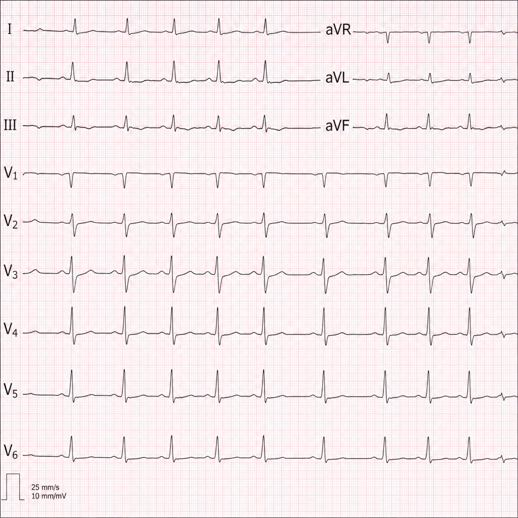
Explanation: ***>0.01 IU/mL*** - A serum level of antibodies **greater than 0.01 IU/mL** is generally considered the **minimum protective level** against tetanus. - This threshold indicates sufficient immunity to prevent clinical tetanus, though higher levels offer an increased margin of protection. *>0.5 IU/mL* - While levels **above 0.5 IU/mL** indicate strong, long-lasting protection, they are not the minimum protective threshold. - This level is often considered indicative of **excellent immunity** that may last for many years. *>5 IU/mL* - A level of **5 IU/mL** or higher signifies a very high antibody titer, far exceeding the basic protective level. - This level is not the standard for basic protection and often seen after **recent vaccination** or booster. *>1.0 IU/mL* - Levels **above 1.0 IU/mL** indicate very good protection, but this is a higher threshold than the minimal protective level. - It suggests a robust immune response, but not the absolute lowest concentration required for immunity.
Explanation: ***Hemodialysis***\n - **Digoxin** has a **large volume of distribution (5-7 L/kg)** and is extensively bound to tissue proteins throughout the body.\n - Only a small fraction of digoxin remains in the plasma, making hemodialysis **ineffective** for removing significant amounts of the drug.\n - Hemodialysis is **not recommended** and **not used** as a treatment for digitalis toxicity.\n\n*Fab fragments*\n - **Digoxin-specific antibody fragments** (Digibind/DigiFab) directly bind to and neutralize free digoxin molecules.\n - This is the **gold standard treatment** for severe digitalis toxicity, especially with life-threatening arrhythmias or significant hyperkalemia.\n - Works rapidly to reverse toxic effects by binding digoxin in the serum.\n\n*Lignocaine*\n - **Lignocaine** (lidocaine) is a class IB antiarrhythmic used to treat **ventricular arrhythmias** caused by digitalis toxicity [1].\n - Suppresses ectopic ventricular activity without further depressing AV conduction.\n - Preferred over other antiarrhythmics like phenytoin for digoxin-induced ventricular tachyarrhythmias.\n\n*Potassium*\n - **Context-dependent use**: Potassium is used in digitalis toxicity **only when hypokalemia is present**.\n - **Hypokalemia** increases myocardial sensitivity to digoxin and worsens toxicity, so correction with potassium is therapeutic.\n - **Important contraindication**: Potassium is **contraindicated in acute digoxin overdose with hyperkalemia**, which commonly occurs due to Na-K-ATPase pump inhibition.\n - When appropriately indicated (hypokalemic state), potassium IS used in management of digitalis toxicity.
Explanation: ***Correct: crystalloids+blood transfusion*** - A 30-40% blood volume loss, indicated by **decreased blood pressure** and **decreased urine output**, corresponds to ATLS **Class III hemorrhagic shock**. - Management for Class III shock requires both **intravenous crystalloids** to restore circulatory volume and **blood transfusion** to replace lost red blood cells and improve oxygen-carrying capacity. - The initial approach follows the **3:1 crystalloid replacement rule**, followed by or concurrent with **packed red blood cells** to address ongoing hemorrhage and maintain oxygen delivery. *Incorrect: blood transfusion alone* - While blood transfusion is crucial for Class III hemorrhagic shock, administering it **alone** without initial crystalloid resuscitation may not adequately address the immediate need for **intravascular volume expansion**. - **Crystalloids** are typically administered first or concurrently to rapidly restore circulating volume and support perfusion before packed red blood cells can be prepared and transfused. *Incorrect: crystalloids infusion* - **Crystalloids alone** would be insufficient for Class III hemorrhage as the patient has experienced significant **red blood cell loss** (30-40% circulating volume) which requires direct replacement to improve oxygen delivery. - While initial crystalloid resuscitation is vital, continuing with crystalloids alone will lead to **dilutional coagulopathy** and failure to correct oxygen-carrying capacity. *Incorrect: plasma therapy* - **Plasma therapy** (e.g., fresh frozen plasma) is primarily used for the correction of **coagulopathy** in actively bleeding patients or those with anticipated massive transfusion. - Although it may be part of a massive transfusion protocol for severe hemorrhage, it is not the primary or sole initial treatment strategy for volume resuscitation and red blood cell replacement in Class III shock.
Explanation: ***PNH*** - Paroxysmal nocturnal hemoglobinuria (PNH) is caused by a defect in the **GPI anchor**, leading to increased susceptibility of red blood cells to lysis by complement [1]. - The condition is characterized by **intracorpuscular defects**, resulting in hemolysis due to the inability to protect red blood cells from complement-mediated destruction [1]. *Portal hypertension* - This condition primarily affects the **portal venous system** and is not directly related to **intracorpuscular defects** in red blood cells. - It commonly leads to complications like **variceal bleeding** and ascites, rather than hemolysis. *PCH* - Paroxysmal cold hemoglobinuria (PCH) involves **cold agglutinins** and triggers hemolysis upon exposure to cold, unrelated to **intracorpuscular defects**. - PCH has a different mechanism involving **IgG antibodies**, resulting in hemolysis when exposed to low temperatures. *Uremic syndrome* - Uremic syndrome is a complication of **chronic kidney disease**, leading to hemolysis but due to **extracorpuscular factors** like toxic metabolites rather than intrinsic defects in red blood cells. - It does not specifically cause **intracorpuscular defects** in hemolysis as seen in PNH. **References:** [1] Kumar V, Abbas AK, et al.. Robbins and Cotran Pathologic Basis of Disease. 9th ed. Red Blood Cell and Bleeding Disorders, pp. 650-651.
Explanation: ***Correct: The free concentration of drug X in blood is higher, facilitating tissue distribution.*** - This is the **MOST accurate and complete** answer because it directly addresses the pharmacokinetic mechanism - Drug X has **lower affinity for albumin** → larger proportion remains **unbound (free)** in plasma - Only **free (unbound) drug** can cross capillary membranes to distribute into tissues - This statement precisely explains both the **cause** (higher free concentration) and **effect** (facilitating tissue distribution) *Drug X will be more available in tissues* - This statement is **factually true** and follows logically from drug X's lower protein binding - However, it's **less precise** than the correct answer because it doesn't explicitly explain the **mechanism** (higher free concentration) - The term "available" is less specific than "free concentration," which is the key pharmacokinetic parameter *Drug Y will be less available in tissues* - This statement is also **factually true** - drug Y's **150× higher albumin affinity** means more drug is bound - Higher protein binding → **smaller free fraction** → less tissue distribution - However, like option 1, this doesn't explicitly state the **mechanistic principle** involving free drug concentration - The question asks for the MOST accurate statement, and this focuses on drug Y rather than explaining the core concept *Toxicity of drug Y may be influenced by multiple factors, not just its binding* - While this is a **true general principle**, it's **not directly relevant** to the specific question - This statement doesn't address the **pharmacokinetic implications** of differential albumin binding - It's too vague and doesn't demonstrate understanding of the relationship between protein binding and tissue distribution - The question specifically asks about the affinity differences and their consequences
Explanation: ***10% sodium chloride*** - **Sodium chloride solution** (common salt) is the **standard preservative** for viscera in toxicological analysis in forensic medicine. - A **saturated solution of sodium chloride** (approximately 26-36%) is ideal, but even 10% solution provides **antimicrobial properties** that prevent putrefaction. - It does **not interfere with chemical tests** for poisons, drugs, or toxins, making it superior for toxicological analysis. - Preserves tissue integrity while allowing accurate detection of volatile and non-volatile poisons. *20% alcohol* - While **rectified spirit (90-95% alcohol)** is used for preserving **blood and urine** samples, dilute alcohol (20%) is **not recommended for viscera**. - Dilute alcohol lacks sufficient antimicrobial strength and may cause tissue shrinkage. - Not the standard choice in forensic toxicology protocols for visceral organs. *10% formalin* - **Formalin** is excellent for **histopathological preservation** but is **contraindicated in toxicological analysis**. - It can **destroy or alter poisons** and interfere with chemical detection methods. - Cross-linking of proteins makes toxin extraction difficult. *Common bile salt* - **Bile salts** are biological detergents with no preservative properties. - Not used in forensic medicine for sample preservation. - No antimicrobial or tissue-stabilizing properties.
Explanation: ***Clearance*** - **Clearance (CL)** is the primary and direct determinant of drug elimination after IV administration. - It represents the **volume of plasma cleared of drug per unit time** (e.g., mL/min or L/hr). - The **rate of elimination** is directly calculated as: Rate = CL × Plasma concentration - Clearance integrates the efficiency of all eliminating organs (liver, kidneys) and is the key parameter determining how fast a drug is removed from the body. - Formula: **CL = Rate of elimination / Plasma concentration** *Lipid solubility* - Lipid solubility affects drug **distribution** and **renal reabsorption** but does not directly determine the rate of elimination. - Highly lipid-soluble drugs may be reabsorbed in renal tubules, but the elimination rate is still governed by clearance. - Lipid solubility is more relevant to drug distribution and metabolism pathways than to the rate of elimination itself. *Volume of distribution* - Volume of distribution (Vd) describes how extensively a drug distributes into tissues versus plasma. - While Vd affects the **half-life** (t½ = 0.693 × Vd/CL), it does NOT directly determine the elimination rate. - A large Vd means more drug in tissues, which affects how long elimination takes, but the actual rate of elimination is still determined by clearance. - Vd is a distribution parameter, not an elimination parameter. *All of the options* - This is incorrect because only **clearance** directly determines the rate of drug elimination. - While lipid solubility and volume of distribution can indirectly influence how long a drug remains in the body, they do not determine the elimination rate itself—clearance does.
Explanation: ***Surgical removal of nasal polyps*** - Septoplasty is a surgical procedure specifically designed to correct a **deviated nasal septum** by repositioning or removing obstructing cartilage and bone. - **Nasal polyps** arise from the mucosa of the nasal cavity or sinuses and require a separate procedure, typically **functional endoscopic sinus surgery (FESS)** or polypectomy. - While septoplasty and polypectomy may sometimes be performed together, polyp removal is **not part of standard septoplasty**. *Submucosal resection of deviated cartilage* - This is the **core component of septoplasty** - removing or repositioning deviated septal cartilage while preserving the mucosal lining. - The submucosal approach maintains structural support while correcting the deviation. *Throat pack* - A **throat pack** is routinely placed during septoplasty to **prevent aspiration of blood and secretions** into the pharynx and esophagus. - It protects the airway and is removed at the end of the procedure. *Nasal packing at the end of surgery* - **Nasal packing** (splints or packs) is commonly placed after septoplasty to **control bleeding, support the septum, and prevent hematoma formation**. - Modern techniques may use absorbable or non-absorbable packing materials.
Explanation: ***Hypocalcemia*** - **Hypocalcemia** prolongs the **repolarization phase** of the action potential in cardiac myocytes, leading to a lengthened **QT interval** on an electrocardiogram. - This increased duration of repolarization places the heart at higher risk for **Torsades de Pointes** and other life-threatening arrhythmias [2], [3]. *Hypernatremia* - **Hypernatremia** primarily affects neurological function and can cause symptoms like **confusion** and **seizures**. - It does not typically lead to a **prolonged QT interval**; instead, it can sometimes be associated with a shortened QT interval or other non-specific ECG changes. *Hyperkalemia* - **Hyperkalemia** primarily causes peaked T waves, a widened QRS complex, and eventually **bradycardia** and **asystole** [1]. - While it drastically alters cardiac conduction, it typically **shortens** rather than prolongs the QT interval. *Hyponatremia* - **Hyponatremia** is associated with cerebral edema and neurological symptoms such as **headaches**, **nausea**, and **altered mental status**. - It generally does not cause a **prolonged QT interval**; significant hyponatremia can sometimes be associated with non-specific ECG changes [1] but not a specific lengthening of the QT interval.
Explanation: ***Low serum calcium*** - **Osteomalacia** is characterized by defective **bone mineralization**, often due to vitamin D deficiency, which leads to impaired calcium absorption and subsequent **hypocalcemia** [1]. - To compensate for low serum calcium, **parathyroid hormone (PTH)** levels increase, further contributing to altered bone metabolism [1]. *Low serum phosphate* - While osteomalacia often features **hypophosphatemia**, it is not always the primary or most consistent finding, as **calcium** dysregulation is central to the disease [1]. - **Secondary hyperparathyroidism** in some cases of osteomalacia can lead to increased phosphate excretion, causing low phosphate, but **hypocalcemia** is more directly related to the mineralization defect [1]. *Normal level of 1, 25 di-hydroxy vit D3* - **1,25-dihydroxyvitami n D3** (calcitriol) levels are typically **low** in nutritional osteomalacia, as this is the active form crucial for calcium absorption and bone mineralization [1]. - Normal levels would contradict the underlying pathology of vitamin D deficiency that causes the condition. *Normal hydroxy proline levels in urine* - **Hydroxyproline** is an amino acid found in collagen, and its urinary excretion reflects **bone turnover**; in osteomalacia, due to defective mineralization and sometimes increased bone resorption, hydroxyproline levels can be elevated or altered. - Normal levels would not be expected given the significant bone metabolic disturbances in osteomalacia.
Get full access to all questions, explanations, and performance tracking.
Start For Free