Anti-TNF alpha drugs are used for the treatment of all of the following diseases except?
Morbidly obese patients with obstructive sleep apnea often experience which of the following conditions, except?
All of the following are risk factors for obstructive sleep apnea EXCEPT:
Polysomnography includes all of the following tests except:
Anti-ds DNA antibody is specific for which condition?
Non-erosive arthritis is characteristic of which condition?
Which of the following antibodies correlates with disease activity for Systemic Lupus Erythematosus?
Sleep apnea is defined as a temporary pause in breathing during sleep lasting at least:
An obese, diabetic patient with hypertension, who is also a smoker and currently on anti-hypertensive and oral hypoglycemic drugs, presents with complaints of apnea during sleep. Polysomnography reveals 5 apneic episodes and 1 hypopneic episode per hour. What is the best next line of management?
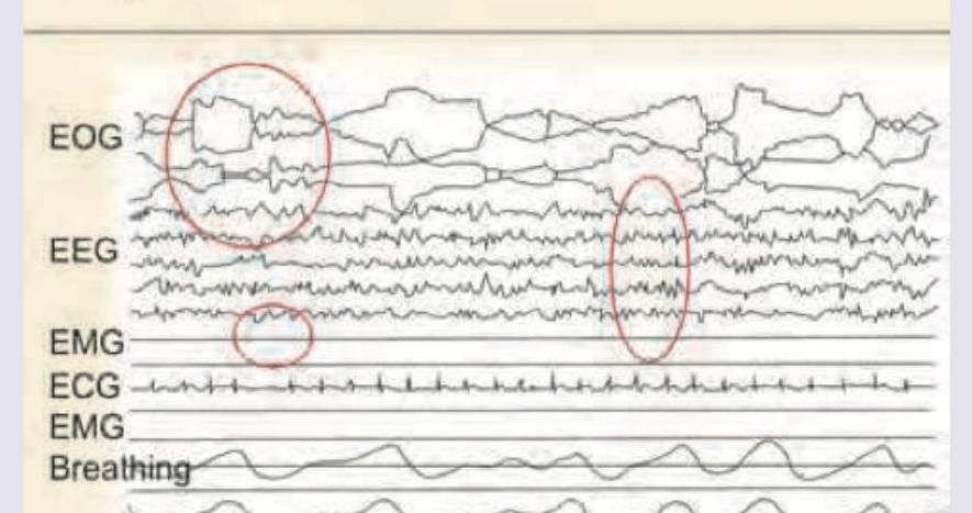
Identify the sleep stage in the following Polysomnograph.

Explanation: Anti-TNF alpha agents (e.g., Infliximab, Etanercept, Adalimumab) are potent biological DMARDs used to treat chronic inflammatory conditions driven by Tumor Necrosis Factor-alpha [1]. **Why Systemic Lupus Erythematosus (SLE) is the correct answer:** Anti-TNF alpha drugs are generally **avoided in SLE** because they can paradoxically induce the formation of autoantibodies (like ANA and anti-dsDNA) [2]. In some patients, these drugs can trigger **Drug-Induced Lupus Erythematosus (DILE)**. While they are occasionally researched for refractory lupus nephritis, they are not a standard treatment and are contraindicated due to the risk of exacerbating the underlying autoimmune process. **Why the other options are incorrect:** * **Seronegative Arthritis (e.g., Ankylosing Spondylitis):** TNF-alpha is a key mediator in the pathogenesis of spinal inflammation. Anti-TNF agents are the first-line biological therapy when NSAIDs fail. * **Psoriatic Arthritis:** These drugs are highly effective in treating both the skin manifestations (psoriasis) and the joint destruction associated with this condition. * **Crohn’s Disease:** TNF-alpha plays a central role in intestinal mucosal inflammation [3]. Agents like Infliximab are gold-standard treatments for inducing and maintaining remission in moderate-to-severe or fistulizing Crohn’s disease. **High-Yield Clinical Pearls for NEET-PG:** 1. **Pre-treatment Screening:** Always screen for **Latent Tuberculosis** (via TST or IGRA) and **Hepatitis B** before starting Anti-TNF therapy, as these drugs can cause reactivation. 2. **Contraindications:** Avoid Anti-TNF drugs in patients with **NYHA Class III/IV Heart Failure** and **Demyelinating diseases** (like Multiple Sclerosis). 3. **Side Effects:** Increased risk of lymphomas and serious opportunistic infections [1].
Explanation: The question asks for the condition **not** typically associated with Obstructive Sleep Apnea (OSA) in morbidly obese patients. While OSA is a major risk factor for cardiovascular disease, the primary hemodynamic consequence is **Right Ventricular Failure (Cor Pulmonale)**, not Left Ventricular Failure (LVF). **1. Why "Left Ventricular Failure" is the correct choice (The Exception):** In OSA, repetitive upper airway collapse leads to nocturnal hypoxemia and hypercapnia. This triggers **pulmonary vasoconstriction**, leading to pulmonary hypertension and subsequent **Right Ventricular (RV) hypertrophy and failure** [2]. While OSA is associated with systemic hypertension (a risk factor for LVF), the direct, hallmark pathophysiological progression in obese OSA patients is toward the right side of the heart. Therefore, LVF is the "odd one out" compared to the direct respiratory-driven effects. **2. Analysis of Incorrect Options:** * **Hypoxemia (B):** Apneic episodes lead to a cessation of airflow, causing a characteristic drop in arterial oxygen saturation ($SaO_2$). * **Hypercapnia (C):** Lack of ventilation during sleep leads to the retention of $CO_2$ [3]. In morbidly obese patients, this may progress to **Obesity Hypoventilation Syndrome (Pickwickian Syndrome)**, where hypercapnia persists during wakefulness. * **Right Ventricular Failure (A):** Chronic nocturnal hypoxemia causes pulmonary hypertension, which increases the afterload on the right ventricle, eventually leading to failure [2]. **Clinical Pearls for NEET-PG:** * **Gold Standard Diagnosis:** Polysomnography (Sleep Study) [1]. * **Definition:** Apnea-Hypopnea Index (AHI) $\geq$ 5 events/hour with symptoms, or $\geq$ 15 events/hour without symptoms. * **Pickwickian Syndrome Triad:** Obesity (BMI $>30$), Sleep Apnea, and Daytime Hypercapnia ($PaCO_2 >45$ mmHg). * **Treatment of Choice:** Continuous Positive Airway Pressure (CPAP).
Explanation: This question focuses on the **STOP-BANG criteria**, a high-yield screening tool used to identify patients at risk for Obstructive Sleep Apnea (OSA). [1] ### **Explanation of the Correct Answer** **Option A (BMI > 30.3)** is the correct answer because it is a "distractor" value. According to the validated STOP-BANG scoring system, the specific threshold for risk is a **BMI > 35 kg/m²**. While obesity (BMI > 30) is a general risk factor for OSA, the standardized screening criteria used in clinical practice and exams specifically use the cut-off of 35. ### **Analysis of Incorrect Options** The other options are all established components of the **STOP-BANG** acronym: * **Option B (Tiredness):** The "**T**" in STOP stands for **Tiredness**, fatigue, or sleepiness during the daytime. [1] * **Option C (Hypertension):** The "**P**" in STOP stands for **Pressure** (High Blood Pressure). OSA is a secondary cause of hypertension; over 50% of OSA patients are hypertensive. * **Option D (Neck Circumference):** The "**N**" in BANG stands for "**Neck circumference**. The threshold is **> 40 cm (16 inches)**. ### **NEET-PG Clinical Pearls: STOP-BANG Score** To excel in Sleep Medicine questions, memorize the STOP-BANG mnemonic: 1. **S**noring (Loud) 2. **T**iredness (Daytime sleepiness) 3. **O**bserved apnea (Choking/gasping) 4. **P**ressure (Hypertension) 5. **B**ody Mass Index (**> 35 kg/m²**) 6. **A**ge (**> 50 years**) 7. **N**eck circumference (**> 40 cm**) 8. **G**ender (**Male**) * **High-Yield Fact:** Gold standard for diagnosis is **In-laboratory Polysomnography (PSG)**. [1] An Apnea-Hypopnea Index (AHI) **≥ 5** with symptoms or **≥ 15** without symptoms is diagnostic. [1]
Explanation: **Explanation:** Polysomnography (PSG) is the gold-standard diagnostic test for sleep disorders, particularly Obstructive Sleep Apnea (OSA) [1], [2]. It is a multi-parametric study that monitors various physiological functions during sleep to determine sleep stages and respiratory events. **Why "Aerial pCO2 measurement" is the correct answer:** Standard PSG measures respiratory effort (via chest/abdominal belts), airflow (via thermistors/nasal pressure transducers), and oxygen saturation. While **End-tidal CO2 (EtCO2)** or **Transcutaneous CO2** may be used in specific pediatric cases or hypoventilation syndromes, "Aerial pCO2" (measuring CO2 levels in the room air) is not a component of a sleep study as it does not reflect the patient's physiological state. **Analysis of other options:** * **Electroencephalography (EEG):** Essential for identifying sleep stages (N1, N2, N3, and REM) and detecting arousals [1]. * **Electrooculography (EOG):** Records eye movements; it is crucial for identifying the onset of REM (Rapid Eye Movement) sleep. * **Pulse Oximetry:** Continuously monitors SpO2 to detect desaturations associated with apneic or hypopneic events [2]. **High-Yield Clinical Pearls for NEET-PG:** 1. **Components of PSG:** EEG, EOG, EMG (Electromyography - usually chin and legs), ECG, Airflow, Respiratory effort, and Pulse Oximetry. 2. **Apnea-Hypopnea Index (AHI):** The primary metric derived from PSG. * Mild OSA: AHI 5–15 * Moderate OSA: AHI 15–30 * Severe OSA: AHI >30 3. **Multiple Sleep Latency Test (MSLT):** The gold standard for diagnosing **Narcolepsy** (measures how quickly a patient falls asleep in a quiet environment during the day). 4. **Gold Standard Treatment:** CPAP (Continuous Positive Airway Pressure) is the treatment of choice for OSA [2].
Explanation: **Explanation:** **Systemic Lupus Erythematosus (SLE)** is the correct answer because Anti-dsDNA (double-stranded DNA) antibodies are highly specific (approx. 97-100%) for this condition [2]. While **ANA (Antinuclear Antibody)** is the best initial screening test due to its high sensitivity [1], **Anti-dsDNA** and **Anti-Smith (Anti-Sm)** are the confirmatory "specific" markers [1], [2]. Clinically, Anti-dsDNA titers correlate with disease activity, particularly the development of **Lupus Nephritis**. **Analysis of Incorrect Options:** * **Systemic Sclerosis (Diffuse):** This is primarily associated with **Anti-Scl-70 (Anti-topoisomerase I)** antibodies. * **CREST Syndrome (Limited Scleroderma):** The hallmark marker is the **Anti-centromere antibody**. (Mnemonic: **C**REST = **C**entromere). * **Sjogren’s Syndrome:** Characterized by **Anti-Ro (SS-A)** and **Anti-La (SS-B)** antibodies [1]. While these can be seen in SLE (associated with neonatal lupus), they are the primary markers for Sjogren’s. **High-Yield Clinical Pearls for NEET-PG:** * **Most Sensitive Test for SLE:** ANA (Best for screening) [1]. * **Most Specific Tests for SLE:** Anti-dsDNA and Anti-Smith [1], [2]. * **Drug-Induced Lupus:** Associated with **Anti-histone antibodies** (Hydralazine, Procainamide, Isoniazid). * **Neonatal Lupus/Congenital Heart Block:** Strongly associated with **Anti-Ro (SS-A)** antibodies [1]. * **Disease Activity Monitoring:** Anti-dsDNA levels and **Complement levels (C3, C4)** are used to monitor flares; as Anti-dsDNA rises, C3/C4 levels typically fall [2].
Explanation: Explanation: The hallmark of joint involvement in Systemic Lupus Erythematosus (SLE) is non-erosive arthritis [1]. Unlike inflammatory arthritides that destroy bone and cartilage, SLE involves synovial inflammation that leads to laxity of ligaments and tendons rather than bony destruction. While patients may present with significant deformities (such as Jaccoud’s arthropathy, characterized by ulnar deviation and swan-neck deformities), these are typically reducible because the underlying joint surface remains intact on X-ray. Analysis of Incorrect Options: * Rheumatoid Arthritis (RA): This is the prototype of erosive arthritis [2]. Chronic synovial pannus formation leads to marginal bony erosions and joint space narrowing, resulting in permanent, non-reducible deformities. * Psoriasis (Psoriatic Arthritis): Characterized by aggressive bone involvement. Radiographic features include "pencil-in-cup" deformities and marginal erosions, often leading to joint destruction (Arthritis mutilans) [1]. * Gout: Chronic tophaceous gout causes "punched-out" erosions with overhanging edges (Martel’s sign) due to the deposition of monosodium urate crystals in and around the joint. NEET-PG High-Yield Pearls: * Jaccoud’s Arthropathy: Classic "reversible" deformity seen in SLE (and Rheumatic Fever). * Most common joint involved in SLE: Small joints of the hands (PIP, MCP) and wrists, usually symmetrical [1]. * X-ray finding in SLE: Soft tissue swelling and periarticular osteopenia, but no marginal erosions. * Synovial fluid in SLE: Usually mildly inflammatory (WBC count <2000–5000/mm³).
Explanation: In Systemic Lupus Erythematosus (SLE), **Anti-dsDNA antibodies** are highly specific and serve as a crucial biomarker for monitoring the disease. The titers of Anti-dsDNA fluctuate in tandem with disease activity; rising levels often precede a clinical flare, particularly **Lupus Nephritis** [1]. Furthermore, these antibodies are directly involved in pathogenesis by forming immune complexes that deposit in the glomerular basement membrane. **Analysis of Options:** * **Anti-Smith (Sm) antibody:** This is the **most specific** antibody for SLE (included in ACR criteria), but its levels remain constant regardless of disease state. It does not correlate with activity or flares. * **Anti-Histone antibody:** This is the hallmark of **Drug-Induced Lupus** (e.g., caused by Hydralazine, Procainamide, or Isoniazid). It is rarely seen in idiopathic SLE. * **Anti-Ro (SS-A) antibody:** Associated with Neonatal Lupus (congenital heart block), Subacute Cutaneous Lupus, and Sjögren’s syndrome. It does not track with SLE disease activity. **NEET-PG High-Yield Pearls:** * **Best Screening Test:** ANA (High sensitivity, low specificity). * **Most Specific Test:** Anti-Smith. * **Best Marker for Disease Activity/Nephritis:** Anti-dsDNA and **low Complement levels (C3, C4)** [1]. * **Drug-Induced Lupus:** Characterized by Anti-Histone (+) and usually Anti-dsDNA (-); it rarely involves the CNS or Kidneys.
Explanation: **Explanation:** **1. Why Option D is Correct:** In clinical sleep medicine, an **apnea** is defined as a complete or near-complete (≥90%) cessation of airflow at the nose and mouth for a duration of **at least 10 seconds** [1]. This 10-second threshold is the standardized diagnostic criterion used in Polysomnography (PSG) to identify clinically significant respiratory events. These pauses are often associated with oxygen desaturation or cortical arousals, leading to fragmented sleep [2]. **2. Why Other Options are Incorrect:** * **Options A (40s) and B (30s):** While apneic episodes can certainly last this long (and are often more severe when they do), they are not the *minimum* requirement for diagnosis. Using these as a baseline would significantly under-diagnose patients with Obstructive Sleep Apnea (OSA). * **Option C (20s):** This is a common distractor. While 20 seconds is a threshold often used in **neonatal medicine** to define apnea of prematurity (or shorter if accompanied by bradycardia/cyanosis), the standard adult definition remains 10 seconds. **3. High-Yield Clinical Pearls for NEET-PG:** * **Apnea-Hypopnea Index (AHI):** This is the gold standard for grading OSA severity [2]. It is calculated as the (Total number of Apneas + Hypopneas) / Total Sleep Time (in hours). * *Mild:* 5–15 events/hr * *Moderate:* 15–30 events/hr * *Severe:* >30 events/hr * **Hypopnea:** Defined as a ≥30% reduction in airflow for ≥10 seconds, accompanied by ≥3% oxygen desaturation or an arousal. * **Gold Standard Investigation:** Polysomnography (Level 1 study). * **Treatment of Choice:** Continuous Positive Airway Pressure (CPAP) [2]. * **Common Association:** Pickwickian Syndrome (Obesity Hypoventilation Syndrome) characterized by obesity, daytime hypercapnia, and sleep-disordered breathing.
Explanation: ### Explanation The patient presents with symptoms suggestive of Obstructive Sleep Apnea (OSA). However, the diagnosis and management of OSA are strictly guided by the **Apnea-Hypopnea Index (AHI)**, which is the number of apnea and hypopnea episodes per hour of sleep [1]. **1. Why "Diet and weight reduction" is correct:** In this case, the patient’s AHI is **6** (5 apneas + 1 hypopnea). According to clinical guidelines: * **Normal:** AHI < 5 * **Mild OSA:** AHI 5–15 * **Moderate OSA:** AHI 15–30 * **Severe OSA:** AHI > 30 Since the patient falls into the **Mild OSA** category and possesses multiple reversible risk factors (obesity, smoking), the initial management is **conservative lifestyle modification** [1]. Weight loss is the most effective non-device intervention to reduce pharyngeal collapsibility and improve AHI [3]. **2. Why other options are incorrect:** * **Nasal CPAP:** This is the "Gold Standard" treatment for OSA [1], but it is typically indicated for **Moderate to Severe OSA** (AHI > 15) or Mild OSA (AHI 5–15) only if the patient has significant symptoms (daytime sleepiness) or co-morbidities like cardiovascular disease [2]. * **Uvulopharyngopalatoplasty (UPPP):** This is a surgical intervention reserved for patients who fail CPAP or have specific anatomical obstructions. It is not a first-line treatment. * **Mandibular sling/Advancement Devices:** These are oral appliances used for mild-to-moderate OSA in patients who cannot tolerate CPAP, but they follow lifestyle modifications. **Clinical Pearls for NEET-PG:** * **Gold Standard Diagnosis:** Overnight Polysomnography. * **Gold Standard Treatment:** Nasal CPAP (Continuous Positive Airway Pressure) [2]. * **Pickwickian Syndrome:** Obesity Hypoventilation Syndrome (BMI > 30 + daytime hypercapnia PaCO2 > 45 mmHg). * **Mallampati Score:** Used to assess the airway; high scores (III and IV) are associated with an increased risk of OSA.
Explanation: ***NREM stage 2*** - The **EEG** shows prominent **sleep spindles** (bursts of 12-14 Hz waves) and **K-complexes** (high-amplitude biphasic waves), which are characteristic features of NREM stage 2 sleep. - The EOG channels indicate slow eye movements or an absence of rapid eye movements, consistent with NREM sleep, while the **EMG shows moderate muscle tone**, higher than in REM sleep but lower than wakefulness. *NREM stage 3* - This stage is characterized by **delta waves**, which are slow waves with high amplitude (0.5-2 Hz, often >75 μV) on the EEG, comprising 20% or more of the epoch, and are not significantly visible here. - While muscle tone is still present, the EEG would primarily show widespread **slow-wave activity**, distinguishing it from the sleep spindles and K-complexes seen in the image. *REM* - **Rapid eye movements** would be clearly visible on the EOG channels, which are not prevalent in this polysomnograph. - The **EMG would show very low muscle tone** (atonia), which is not the case here, and the EEG would largely consist of low-voltage, mixed-frequency activity, similar to wakefulness. *NREM stage 1* - This stage is typically characterized by a **disappearance of alpha waves** from the EEG and the presence of **theta waves** (4-7 Hz). - While there may be slow eye movements on the EOG, **sleep spindles and K-complexes are absent** in NREM stage 1, making it distinct from the presented polysomnograph.
Normal Sleep Physiology
Practice Questions
Sleep-Disordered Breathing
Practice Questions
Insomnia
Practice Questions
Hypersomnias
Practice Questions
Circadian Rhythm Disorders
Practice Questions
Parasomnias
Practice Questions
Sleep-Related Movement Disorders
Practice Questions
Sleep in Medical Disorders
Practice Questions
Sleep in Psychiatric Disorders
Practice Questions
Sleep Diagnostics
Practice Questions
Pharmacologic Management of Sleep Disorders
Practice Questions
Non-pharmacologic Sleep Interventions
Practice Questions
Get full access to all questions, explanations, and performance tracking.
Start For Free