Which of the following is a feature of Vit B12 deficiency anemia?
Which parameter is primarily used to diagnose macrocytosis in a complete blood count (CBC)?
Earliest manifestation of megaloblastic anemia is
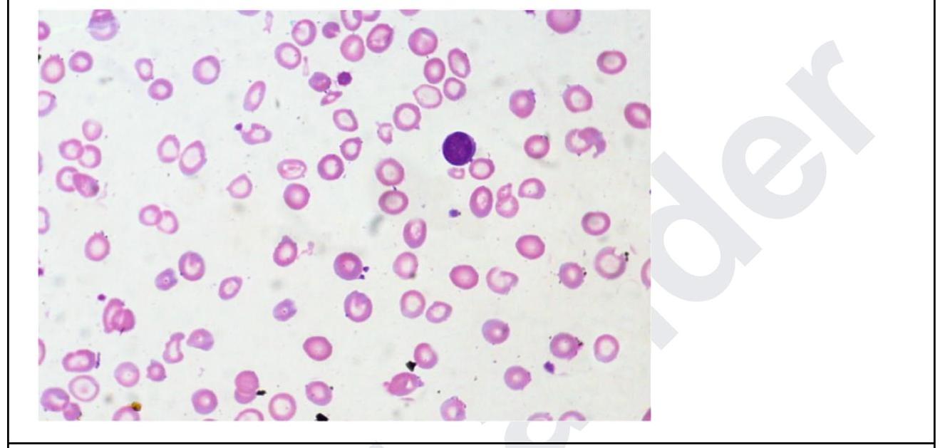
A 35 year old woman presents with fatigue. Investigations revealed the following: Hb, 5 g/dL; MCH, 24; low MCV; leukocytes, 11,000/ uL, and platelets, 5 lakhs. The peripheral smear is shown below. What is the diagnosis? Normal values: - Mean cell volume (MCV); 90 ± 8 fL - Mean cell Hb(MCH); 30 ± 3 pg

Anemia with reticulocytosis is seen in -
A patient has MCV <80, MCH <23. Which type of anaemia shall be classified?
What is a known complication of Parvovirus B19 infection?
Which of the following is not likely to be seen in a patient with Paroxysmal Nocturnal Hemoglobinuria (PNH)?
Bleeding crisis in acute idiopathic thrombo-cytopenic purpura is managed by all except -
A 40-year-old presents with cirrhosis and low ferritin. What is the next step in management?
Explanation: **All of the options** - **Vitamin B12 deficiency anemia** is a type of **megaloblastic anemia** characterized by impaired DNA synthesis, leading to large, immature red blood cells and neutrophils [1]. - The presence of **macro-ovalocytes** and **hypersegmented neutrophils** are classic hematological features seen on a peripheral blood smear [1]. *Macro-ovalocytes* - **Macro-ovalocytes** are abnormally large, oval-shaped red blood cells, which result from arrested maturation due to the deficiency. - While a hallmark of B12 deficiency, it is not the sole identifying feature, as other megaloblastic anemias can also present with them [2]. *Megaloblastic anemia* - **Megaloblastic anemia** is a broad category of anemia characterized by large, immature, and dysfunctional red blood cells, which is the primary classification for B12 deficiency [2]. - While accurate, it doesn't encompass the specific morphological findings seen in the blood smear of B12 deficiency, unlike the other options. *Hypersegmented neutrophils* - **Hypersegmented neutrophils** are neutrophils with five or more nuclear lobes, a characteristic sign of impaired DNA synthesis affecting granulopoiesis [1]. - This feature is highly specific to **megaloblastic anemias**, distinguishing them from other causes of macrocytosis.
Explanation: ***Mean Corpuscular Volume (MCV)*** - **MCV** measures the **average volume of red blood cells**, making it the primary indicator for classifying them as microcytic, normocytic, or macrocytic. - An **elevated MCV** (typically above 100 fL) indicates **macrocytosis**, meaning the red blood cells are larger than normal. *Mean Corpuscular Hemoglobin Concentration (MCHC)* - **MCHC** reflects the **average concentration of hemoglobin** in red blood cells, primarily used to classify cells as normochromic or hypochromic. - It does **not directly measure cell size** and therefore is not used to diagnose macrocytosis. *Hematocrit* - **Hematocrit** measures the **percentage of red blood cells** in a given volume of blood. - While it indicates the overall red cell mass, it **does not provide information about the average size** of individual red blood cells. *None of the options* - As **MCV** is explicitly listed and is the correct parameter for diagnosing macrocytosis, this option is incorrect. - The other parameters listed are important for other aspects of red blood cell analysis but not for classifying cell size.
Explanation: ***Hypersegmented neutrophils*** - The earliest manifestation of megaloblastic anemia includes the presence of **hypersegmented neutrophils**, which have more than five lobes in their nuclei [1][2]. - This finding is indicative of impaired DNA synthesis often associated with **vitamin B12** or **folate deficiency**. *Basophilic stippling* - **Basophilic stippling** is more commonly linked to lead poisoning and certain alcohol-related disorders rather than megaloblastic anemia. - It reflects RNA aggregate remnants in red blood cells, which is not a primary feature of this type of anemia. *Cabot ring* - **Cabot rings** are seen in conditions like **pernicious anemia** but are not the **earliest manifestation**; they are infrequently encountered. - This abnormality is related to nuclear remnant material and lacks direct correlation to megaloblastic changes. *Macrocytosis* - **Macrocytosis** refers to the increased size of red blood cells and can be found in megaloblastic anemia but is not the **initial manifestation**. - It may develop later as the anemia progresses, whereas hypersegmented neutrophils appear much earlier. **References:** [1] Cross SS. Underwood's Pathology: A Clinical Approach. 6th ed. Common Clinical Problems From Blood And Bone Marrow Disease, pp. 593-594. [2] Kumar V, Abbas AK, et al.. Robbins and Cotran Pathologic Basis of Disease. 9th ed. Red Blood Cell and Bleeding Disorders, p. 654.
Explanation: ***Iron-deficiency anemia*** - The patient presents with **severe anemia (Hb 5 g/dL)**, **low MCH (24 pg)**, and **low MCV**, which are classic indicators of **microcytic, hypochromic anemia** [3]. The peripheral smear shows **hypochromic microcytic red cells** with abundant central pallor and **anisopoikilocytosis**, consistent with iron-deficiency anemia [1]. - While the **platelet count is elevated (5 lakhs)**, it can occur in iron deficiency as reactive thrombocytosis [1]. Leukocytosis in the absence of infection may be a mild reactive process secondary to severe anemia. *Essential thrombocytosis* - This is a **myeloproliferative neoplasm** characterized by significantly elevated platelet counts (often > 450,000/uL), but typically does not present with severe anemia, low MCH, or low MCV. - The primary issue in this patient is severe anemia with microcytic hypochromic features, not isolated thrombocytosis. *Thalassemia major* - While thalassemia major also presents with **microcytic, hypochromic anemia** and can have a very low MCV, it usually manifests in early childhood and is associated with significant **hemolysis**, **splenomegaly**, and characteristic red blood cell morphology such as **target cells** and **nucleated red blood cells** [2]. - The extremely low Hb and microcytic indices alone are not enough to distinguish it from severe iron deficiency without further specific markers like iron studies or hemoglobin electrophoresis. *Megaloblastic anemia* - Megaloblastic anemia is characterized by **macrocytic anemia** (high MCV), which is the opposite of the low MCV presented in this case. - It typically results from **vitamin B12** or **folate deficiency** and the peripheral smear would show **macro-ovalocytes** and **hypersegmented neutrophils**, which are not seen here. **References:** [1] Cross SS. Underwood's Pathology: A Clinical Approach. 6th ed. Common Clinical Problems From Blood And Bone Marrow Disease, pp. 590-591. [2] Kumar V, Abbas AK, et al.. Robbins and Cotran Pathologic Basis of Disease. 9th ed. Red Blood Cell and Bleeding Disorders, p. 648. [3] Kumar V, Abbas AK, et al.. Robbins and Cotran Pathologic Basis of Disease. 9th ed. Red Blood Cell and Bleeding Disorders, pp. 638-639.
Explanation: ***Hemolysis*** - Reticulocytosis indicates a compensatory response to anemia, often occurring in hemolytic processes where the **bone marrow increases red blood cell production** in response to red blood cell destruction. - Conditions like **sickle cell disease** or **autoimmune hemolytic anemia** lead to hemolysis, further confirming increased reticulocyte count. *Iron deficiency anemia* - Typically presents with a **low reticulocyte count** as the bone marrow does not have sufficient iron to produce new red blood cells. - This condition is characterized by **microcytic, hypochromic** red blood cells due to inadequate iron stores. *Vitamin B12 deficiency* - Often results in a **macrocytic anemia** with a variable reticulocyte count; however, reticulocytosis is generally not seen initially. - This deficiency affects DNA synthesis, leading to ineffective erythropoiesis and the presence of **megaloblastic changes**. *Aplastic anemia* - Characterized by a **decrease in all types of blood cells** (pancytopenia) and typically has a **low reticulocyte count** due to bone marrow failure. - There is insufficient production of red blood cells, hence **reticulocytosis is not observed**.
Explanation: ***Microcytic hypochromic*** - A **Mean Corpuscular Volume (MCV)** less than **80 fL** indicates **microcytosis** (small red blood cells) [1]. - A **Mean Corpuscular Hemoglobin (MCH)** less than **23 pg** indicates **hypochromia** (pale red blood cells due to reduced hemoglobin content) [1]. *Normocytic normochromic* - This classification refers to red blood cells with **normal MCV (80-100 fL)** and **normal MCH (27-32 pg)**. - Examples include anemia of chronic disease or acute blood loss, which do not fit the given lab values. *Normocytic hypochromic* - While **hypochromia (MCH <23)** is present, the **MCV is less than 80 fL**, which makes it microcytic, not normocytic. - This combination is not a standard classification; hypochromia typically accompanies microcytosis [1]. *Hyperchromic macrocytic* - **Macrocytic anemia** is characterized by an **MCV >100 fL**, which is the opposite of the given MCV of <80. - The term "hyperchromic" is generally not used for anemia classification because red blood cells have a maximal hemoglobin concentration and cannot be truly hyperchromic. **References:** [1] Cross SS. Underwood's Pathology: A Clinical Approach. 6th ed. Common Clinical Problems From Blood And Bone Marrow Disease, pp. 590-591.
Explanation: Aplastic anemia - Parvovirus B19 has a tropism for erythroid progenitor cells in the bone marrow, specifically targeting and destroying them [1]. - This destruction can lead to a transient aplastic crisis, especially in individuals with pre-existing hemolytic conditions, causing a severe drop in red blood cell production [1]. Erythema infectiosum - This is the most common clinical manifestation of Parvovirus B19 infection, also known as fifth disease, characterized by a "slapped cheek" rash [1]. - While it is a symptom or disease caused by the virus, it is not considered a complication in the sense of a secondary, adverse outcome. Arthritis - Arthralgia and arthritis are common manifestations of Parvovirus B19 infection, particularly in adults, especially women [1]. - Similar to erythema infectiosum, it is a direct clinical manifestation rather than a "complication" representing a secondary, undesirable event. All of the options - While erythema infectiosum and arthritis are common clinical presentations of Parvovirus B19, they are direct disease manifestations. - Aplastic anemia stands out as a true complication, representing a secondary and potentially severe adverse outcome due to the virus's specific cellular tropism [1].
Explanation: Leukemia - While PNH can transform into **acute myeloid leukemia (AML)** in a small percentage of cases, it is not a common or direct presentation, making it the *least likely* immediate finding among the options. - The primary pathophysiology of PNH involves a defect in hematopoietic stem cells leading to complement-mediated destruction, not malignant proliferation of myeloid or lymphoid cells as seen in leukemia. *Thrombosis* - **Thrombosis** is a major cause of morbidity and mortality in PNH, occurring due to complement activation and platelet activation on the surface of GPI-deficient cells. - It most commonly affects unusual sites like the **hepatic** or **mesenteric veins**, and cerebral venous sinuses. *Aplastic anemia* - **Aplastic anemia** is closely associated with PNH, as both with conditions can arise from a defect in hematopoietic stem cells. - PNH clones are often detectable in patients with aplastic anemia, and some cases of PNH evolve from or into aplastic anemia. *Hemolysis* - **Hemolysis** is a hallmark of PNH, caused by the absence of **GPI-anchored proteins (CD55 and CD59)** on red blood cells, making them susceptible to complement-mediated destruction [1]. - This leads to intravascular hemolysis, resulting in characteristic symptoms like **dark urine** (hemoglobinuria), especially in the morning [1].
Explanation: ***Eltrombopag*** - **Eltrombopag** is a **thrombopoietin receptor agonist** used for chronic idiopathic thrombocytopenic purpura (ITP) to increase platelet production. - It is **not** used for the immediate management of an acute bleeding crisis, as its effects on platelet counts take several days to manifest. *Intravenous immunoglobulin* - **Intravenous immunoglobulin (IVIG)** works by blocking **Fc receptors** on macrophages, thereby reducing the destruction of antibody-coated platelets. - It is a **first-line treatment** for acute ITP, especially in cases with severe bleeding or very low platelet counts, providing a rapid increase in platelet count. *Prednisolone* - **Prednisolone**, a corticosteroid, is a **first-line treatment** for acute ITP, as it suppresses the immune system and reduces antibody production and platelet destruction. - It helps to quickly raise platelet counts and is effective in managing bleeding episodes, though its effects are not as immediate as IVIG. *RhIG* - **Rh immune globulin (RhIG)** is used in **Rh-positive** patients with ITP to cause a transient hemolytic anemia, which occupies splenic macrophages and reduces platelet destruction. - It `is an effective option` for acute ITP, particularly in patients who require a rapid increase in platelet count and are Rh-positive.
Explanation: ***Iron supplements*** - **Cirrhosis** can lead to **malnutrition** and **impaired iron absorption**, resulting in **iron deficiency anemia**. [2] - **Low ferritin** in a patient with cirrhosis directly indicates **iron deficiency**, making iron supplementation the appropriate initial step to replete iron stores. [3] *Phlebotomy* - **Phlebotomy** is indicated in conditions of **iron overload**, such as **hemochromatosis**, where ferritin levels would be high, not low. [1] - Doing this in a patient with **iron deficiency** would worsen their anemia and be detrimental. *Liver biopsy* - A **liver biopsy** is typically performed to diagnose the cause or assess the stage of **liver disease**, or to evaluate for **liver masses**. [1] - It is not indicated as the next step specifically for **low ferritin** in cirrhosis, as the cause of low ferritin (iron deficiency) is evident and manageable. *Transfusion* - **Blood transfusion** is reserved for patients with severe, **symptomatic anemia** or acute bleeding, where rapid correction of hemoglobin is necessary. - In a patient with **low ferritin** but no mention of severe anemia symptoms, iron supplementation is the first-line treatment.
Get full access to all questions, explanations, and performance tracking.
Start For Free