
Gastroenterology — MCQs

On this page
Which virus is typically associated with serpiginous ulcers in the distal esophagus?
The differentiating feature between IBS and organic GI disease is:
A 42-year-old patient with obstructive jaundice. Alp, Ggt, haptoglobin all increased. The most likely cause is:
Which of the following statements about nutcracker esophagus is correct?
Which of the following laboratory values is NOT a component of the MELD score?
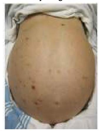
A patient with a history of chronic liver disease presents with abdominal distension, jaundice, and pruritis. Ascitic fluid analysis revealed a neutrophil count >650 per cubic mm. What is the most likely diagnosis?
Which of the following statements is incorrect regarding King's Criteria for acute fulminant liver failure?
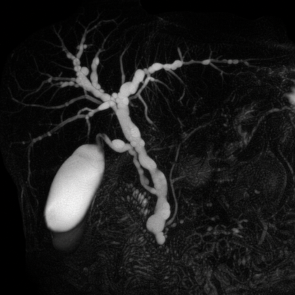
A 35-year-old male presents with recurrent episodes of abdominal pain, jaundice, and fatigue and underwent MRCP. What will be the most likely diagnosis?
Which statement is true regarding Crohn's disease?
Common cause of chronic pancreatitis
Practice by Chapter
Esophageal Disorders
Practice Questions
Peptic Ulcer Disease
Practice Questions
Inflammatory Bowel Disease
Practice Questions
Irritable Bowel Syndrome
Practice Questions
Malabsorption Syndromes
Practice Questions
Pancreatitis (Acute and Chronic)
Practice Questions
Gastrointestinal Bleeding
Practice Questions
Liver Diseases and Cirrhosis
Practice Questions
Viral Hepatitis
Practice Questions
Biliary Tract Disorders
Practice Questions
Gastrointestinal Motility Disorders
Practice Questions
Gastrointestinal Malignancies
Practice Questions
Want unlimited practice?
Get full access to all questions, explanations, and performance tracking.
Scan to download app