
Gastroenterology — MCQs

On this page
Which of the following statements about blind loop syndrome is false?
What is the primary cause of dumping syndrome?
A 14-year-old boy presents with recurrent episodes of hepatitis. Ophthalmoscopic evaluation reveals KF rings and serum ceruloplasmin levels are < 20 mg/dl. What is the treatment of choice for initial therapy?
Which vitamin is not deficient in celiac disease?
Which of the following is not a valid test for investigating fat malabsorption?
A patient comes with abdominal pain, jaundice, and portal hypertension. Anastomosis between which of the following veins is seen?
A 30-year-old male is found to be positive for HBsAg and HBeAg and is diagnosed with chronic hepatitis B. The patient's viral load is 2 × 10^5 IU/mL and ALT is elevated (2× upper limit of normal). What is the appropriate management in this patient?
A patient with a history of alcohol dependence syndrome presents with sudden and unintentional weight loss. What is the most likely diagnosis?
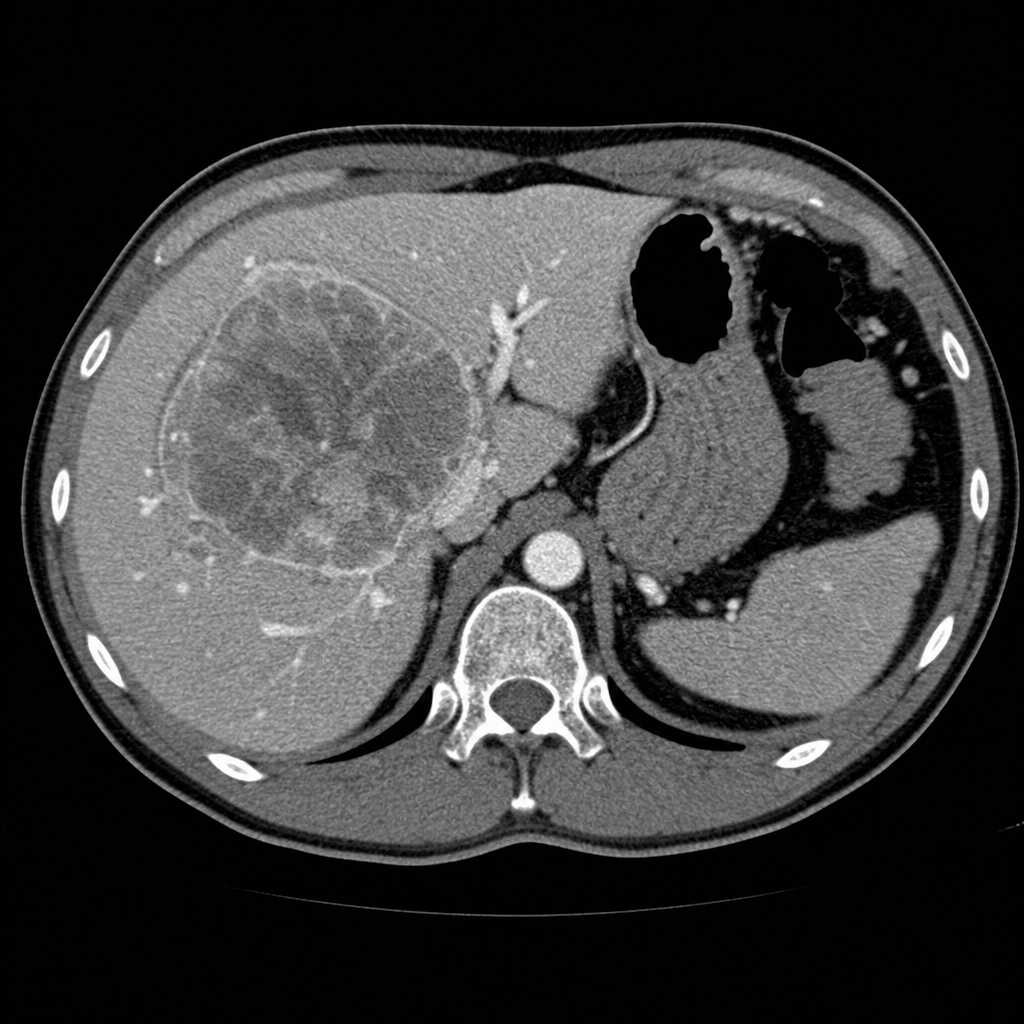
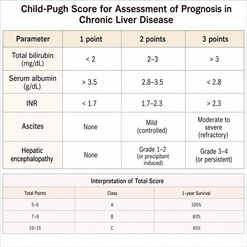
Which of the following criteria is used to assess the prognosis of liver disease?
A 42-year-old patient with obstructive jaundice. Alp, Ggt, haptoglobin all increased. The most likely cause is:
Practice by Chapter
Esophageal Disorders
Practice Questions
Peptic Ulcer Disease
Practice Questions
Inflammatory Bowel Disease
Practice Questions
Irritable Bowel Syndrome
Practice Questions
Malabsorption Syndromes
Practice Questions
Pancreatitis (Acute and Chronic)
Practice Questions
Gastrointestinal Bleeding
Practice Questions
Liver Diseases and Cirrhosis
Practice Questions
Viral Hepatitis
Practice Questions
Biliary Tract Disorders
Practice Questions
Gastrointestinal Motility Disorders
Practice Questions
Gastrointestinal Malignancies
Practice Questions
Want unlimited practice?
Get full access to all questions, explanations, and performance tracking.
Scan to download app