
Gastroenterology — MCQs

On this page
A patient presents with a history of mild diarrhea, blood in stools, and multiple fistulas. What is the most probable diagnosis?
Oral examination is indicated in which of the following conditions?
Which of the following is not a marker of the active replicative phase of chronic hepatitis B?
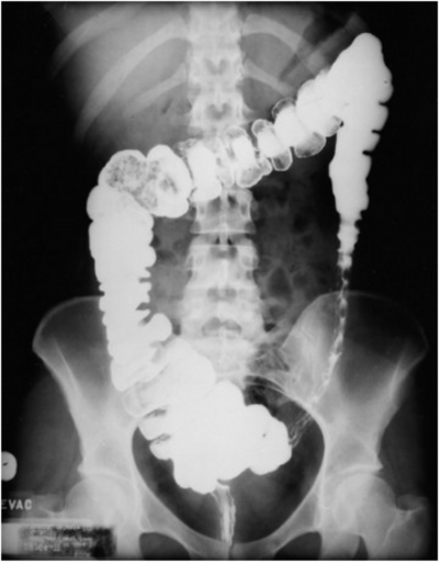
A 85-year-old man presents with severe crampy abdominal pain which is worse after eating, associated with frequent nausea, bloating and watery diarrhoea. His other medical problems include coronary artery disease, hypertension, hypercholesterolemia. He has a 40-pack-year smoking history. Barium enema is as shown. What is the most probable cause?
A 60-year-old man with a known history of hemochromatosis, cirrhosis, and portal hypertension presented to the emergency department with altered mental status. The patient's attendant reported that over the last three days, the patient has become confused. There is no history of melena or hematemesis. For chronic ascites, diet control and spironolactone are being administered regularly. In the past, the patient experienced an episode of variceal bleeding for which he was prescribed propranolol, and no further episodes have occurred. On examination, the patient is not well-oriented to time and place but is oriented to person. He is afebrile, and his vital signs are stable; however, ascites and asterixis are notable. Laboratory investigations show a hemoglobin of 10.1 g/dL, creatinine of 1.4 mg/dL, and blood urea nitrogen of 45 mg/dL. Paracentesis revealed clear fluid with 800 WBC (40% neutrophils). Which statement regarding this condition is false?
All of the following are used in the treatment of acute pancreatitis EXCEPT?
All of the following can predispose to non-cirrhotic portal fibrosis, EXCEPT:
A 40-year-old male with a history of jaundice and ascites is a known alcoholic. Which of the following statements is true regarding his condition?
A 39-year-old female presents with chronic abdominal cramps, watery diarrhea, and periodic facial flushing. Examination reveals wheezing and a slightly enlarged liver. Workup reveals several masses within the liver and a large mass in the small intestine. Which of the following substances is likely to be elevated in her urine?
A 40-year-old male patient presented with mild abdominal pain, mild constipation with a feeling of incomplete evacuation, and mucus in stools for the past four years. On examination, tenderness is present in the left iliac fossa. What is the most likely diagnosis?
Practice by Chapter
Esophageal Disorders
Practice Questions
Peptic Ulcer Disease
Practice Questions
Inflammatory Bowel Disease
Practice Questions
Irritable Bowel Syndrome
Practice Questions
Malabsorption Syndromes
Practice Questions
Pancreatitis (Acute and Chronic)
Practice Questions
Gastrointestinal Bleeding
Practice Questions
Liver Diseases and Cirrhosis
Practice Questions
Viral Hepatitis
Practice Questions
Biliary Tract Disorders
Practice Questions
Gastrointestinal Motility Disorders
Practice Questions
Gastrointestinal Malignancies
Practice Questions
Want unlimited practice?
Get full access to all questions, explanations, and performance tracking.
Scan to download app