
Gastroenterology — MCQs

On this page
All are advantages of Zinc therapy for Wilson disease, except:
A patient with Barrett's esophagus underwent chromoendoscopy. What dye is used in this patient for cancer detection?
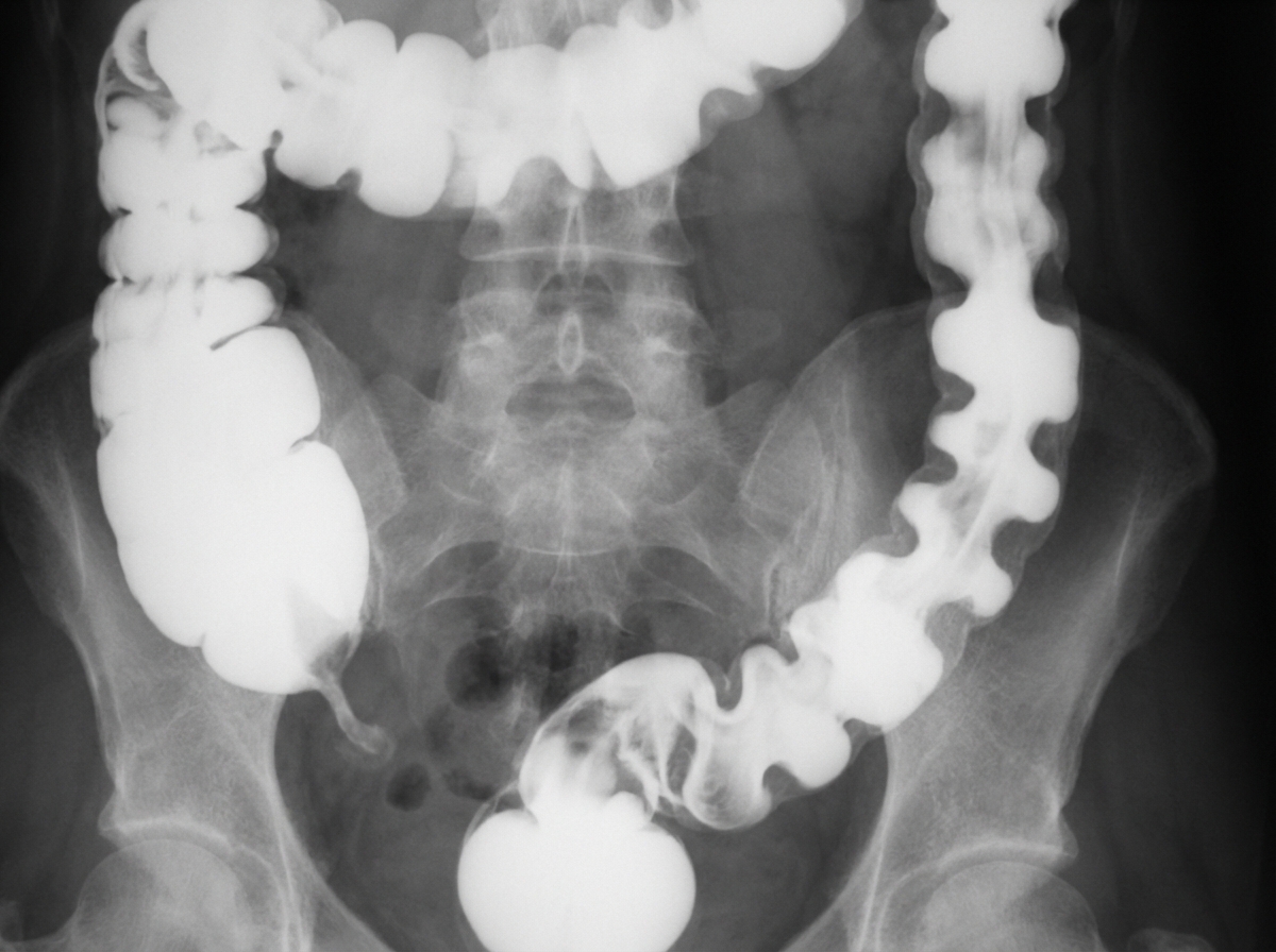
A 70-year-old man presents with severe crampy abdominal pain which is worse after eating. The patient also complains of frequent nausea, bloating, watery diarrhea, and weight loss. His other medical problems include coronary artery disease, hypertension, and hypercholesterolemia. He has a 30-pack-year smoking history. A barium enema is shown. Which of the following statements is true?
Which of the following is NOT part of Charcot's triad?
What is a highly sensitive and specific marker for detecting intestinal inflammation in ulcerative colitis?
Zollinger-Ellison syndrome is not associated with which of the following?
A patient with a history of diabetes mellitus and hypothyroidism presents with passing stools and failure to gain weight. What is the further diagnostic investigation to be done?
A 40-year-old man with a history of Crohn's disease has recently undergone intestinal resection and developed short bowel syndrome as a complication. Which of the following is not a feature of short bowel syndrome?
A patient with steatorrhea has microcytic anemia. The 5-hour urine excretion of D-Xylose after a 25 gm oral load is 2.5 gm. What is the most likely diagnosis?
Ingestion of an osmotically active substance can cause osmotic diarrhea. Which of the following can cause osmotic diarrhea?
Practice by Chapter
Esophageal Disorders
Practice Questions
Peptic Ulcer Disease
Practice Questions
Inflammatory Bowel Disease
Practice Questions
Irritable Bowel Syndrome
Practice Questions
Malabsorption Syndromes
Practice Questions
Pancreatitis (Acute and Chronic)
Practice Questions
Gastrointestinal Bleeding
Practice Questions
Liver Diseases and Cirrhosis
Practice Questions
Viral Hepatitis
Practice Questions
Biliary Tract Disorders
Practice Questions
Gastrointestinal Motility Disorders
Practice Questions
Gastrointestinal Malignancies
Practice Questions
Want unlimited practice?
Get full access to all questions, explanations, and performance tracking.
Scan to download app