Which of the following is a common cause of acute pancreatitis?
Which of the following conditions is a known precipitating factor for hepatic encephalopathy?
All are true regarding familial adenomatous polyposis except which of the following?
A 60-year-old male experienced a sudden fall in the toilet, with a blood pressure of 90/50 mm Hg and a pulse of 100 beats per minute. His relatives reported that his stool was black or dark in color. Further careful history revealed that he was a known case of hypertension and coronary artery disease, and was regularly taking aspirin, atenolol, and isosorbide dinitrate. What is the most likely diagnosis?
Serum amylase is not raised in
A 45-year-old gentleman has undergone truncal vagotomy and pyloroplasty for bleeding duodenal ulcer seven years ago. Now he has intractable recurrent symptoms of peptic ulcer. All of the following suggest the diagnosis of Zollinger-Ellison syndrome, except:
Which of the following statements about Wilson's disease is true?
Absence of the intrahepatic bile ducts leads to which syndrome?
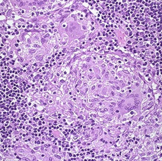
A 40-year-old patient presents with chronic abdominal pain and diarrhea, passing 22-25 stools per day, accompanied by weight loss. On examination, she has mild fever and right lower quadrant tenderness. A lower GI endoscopy reveals the following histopathological findings. An intern in the medicine ward generates a list of differential diagnoses for this patient. Which of the following options is most likely the diagnosis?

Multiple intrahepatic bile duct dilations with bile lakes and concurrent sepsis are suggestive of.
Explanation: ***Gallstones*** - **Gallstones** are the most common cause of acute pancreatitis, as they can **obstruct the common bile duct** at the ampulla of Vater, leading to reflux of bile into the pancreatic duct [1]. - This obstruction causes **pancreatic enzyme activation** within the gland, leading to autodigestion and inflammation [1]. *Elevated serum amylase levels* - **Elevated serum amylase levels** are a diagnostic marker for acute pancreatitis, not a cause. - They indicate pancreatic injury and enzyme release but do not initiate the condition. *Acute alcohol consumption* - **Acute alcohol consumption** is a significant cause of acute pancreatitis but is the second leading cause after gallstones. - While alcohol triggers premature activation of pancreatic enzymes, it is not the *most common* cause. *None of the options* - This option is incorrect because **gallstones** are a well-established and the most common cause of acute pancreatitis [1].
Explanation: **Gastrointestinal bleeding** - **Gastrointestinal bleeding** leads to an increased protein load in the intestines (from blood), which is then broken down by bacteria into **ammonia** [1]. - This elevated ammonia level overwhelms the liver's capacity to detoxify it, leading to its accumulation in the systemic circulation and subsequent entry into the brain, causing **hepatic encephalopathy** [1]. *Increased potassium* - While electrolyte imbalances can affect neurological function, **hyperkalemia** (increased potassium) is not a direct or prominent precipitating factor for hepatic encephalopathy. - Hyperkalemia primarily affects **cardiac and neuromuscular excitability**, rather than ammonia metabolism. *Constipation* - **Constipation** prolongs the transit time of stool in the colon, allowing more time for gut bacteria to produce **ammonia** from protein breakdown [1]. - This increased production of ammonia contributes to its accumulation and can precipitate or worsen hepatic encephalopathy, but it is not the most significant factor like GI bleeding. (The question is which of the following is THE precipitating factor for HE, not the other way around) *Dehydration* - **Dehydration** can lead to **prerenal azotemia** and hemoconcentration, which may increase the blood urea nitrogen (BUN) and worsen renal function. - While dehydration can indirectly affect the body's ability to clear toxins, it is not a direct precipitating factor for hepatic encephalopathy in the same way as increased ammonia production or reduced effective circulating volume.
Explanation: ***Autosomal recessive*** - **Familial adenomatous polyposis (FAP)** is inherited in an **autosomal dominant** pattern, not autosomal recessive. - It is caused by a germline mutation in the **APC gene** on chromosome 5q21-q22. *Associated endocrine involvement can be present* - **Gardner syndrome**, a variant of FAP, is associated with **extracolonic manifestations**, including benign osteomas, desmoid tumors, and epidermal cysts, which can be linked to endocrine disturbances [1]. - Other manifestations can include **adrenal adenomas** and **thyroid cancer**, reflecting potential widespread systemic involvement. *Multiple duodenal polyps* - FAP is characterized by the development of numerous **adenomatous polyps** throughout the gastrointestinal tract, especially in the **colon and rectum**. - **Duodenal polyps**, particularly in the periampullary region, are common and can malignant change, requiring regular surveillance [1]. *More > 100 polyps in the colon* - The defining feature of FAP is the presence of **hundreds to thousands of colorectal adenomatous polyps**, often exceeding 100. - These polyps have a nearly **100% risk of malignant transformation** into colorectal cancer if left untreated.
Explanation: ### Gastric ulcer with hemorrhage - The patient's **hypotension** (90/50 mmHg), **tachycardia** (100 bpm), and a sudden fall are indicative of **hypovolemic shock**, most likely due to significant **blood loss** [1]. - **Black or dark stools (melena)** are a classic sign of **upper gastrointestinal bleeding**, commonly caused by a **gastric ulcer**, especially in a patient taking **aspirin**, which can cause gastrointestinal irritation and ulceration [1]. *Acute myocardial infarction* - While the patient has coronary artery disease, the primary symptoms presented (melena, signs of hypovolemia) are not typical of an **acute myocardial infarction**, which usually causes chest pain, dyspnea, and EKG changes. - Patients with MI can experience hypotension, but it's usually due to **cardiogenic shock**, and the GI bleed evidence points elsewhere. *Stroke* - A stroke typically presents with **focal neurological deficits** (e.g., weakness, speech difficulties), which are not described in this scenario. - While a fall can be a symptom of a stroke, the complete clinical picture including melena strongly points away from a primary neurological event. *Pulmonary embolism* - Pulmonary embolism typically presents with **acute dyspnea**, **chest pain**, and **tachycardia**, often without hypotension initially, or with right-sided heart strain. - The presence of melena and signs of hypovolemic shock are not consistent with a primary diagnosis of **pulmonary embolism**.
Explanation: Ruptured ovarian cyst - A ruptured ovarian cyst involves the *gonads* and does not typically result in the release of **amylase** into the bloodstream. - While it can cause *acute abdominal pain*, it does not directly affect organs that produce significant amounts of amylase, such as the *pancreas* or *salivary glands*. Cholecystitis - **Cholecystitis** can cause *mild elevations* in serum amylase, particularly if there is associated *pancreatic inflammation* [1] or *gallstone pancreatitis* [2]. - Obstruction of the bile duct can lead to reflex pancreatic exocrine dysfunction and increased amylase. Peptic ulcer perforation - A **perforated peptic ulcer** can lead to the release of *gastric or duodenal contents* into the *peritoneal cavity*, causing **chemical peritonitis** [1]. - This irritation can sometimes trigger a *sympathetic response* leading to mild, transient increases in serum amylase or *pancreatic inflammation*. Intestinal ischemia - **Intestinal ischemia** can cause *cellular damage* and *inflammatory responses* within the bowel wall. - In severe cases, this can lead to the release of various enzymes, including *amylase*, from the damaged intestinal tissue or through a *secondary inflammatory effect* on the pancreas.
Explanation: ***Serum gastrin value of 200 pg/ml with secretin stimulation*** - A **positive secretin stimulation test** for Zollinger-Ellison syndrome (ZES) is indicated by a rise in serum gastrin of **≥ 200 pg/mL (or 110 pg/mL depending on the reference range)** above baseline after secretin administration. - A value of 200 pg/ml with secretin stimulation is not diagnostic if the baseline is not known or if the rise from baseline is not significant (i.e., less than 200 pg/ml absolute rise or less than 110 pg/ml rise depending on the specific criteria used). In the context of the other options, this relatively lower value is the *least* indicative of ZES. *Basal acid output of 15 meq/hour* - In a patient with prior vagotomy, a **basal acid output (BAO) of 15 meq/hour** is significantly elevated and highly suggestive of Zollinger-Ellison syndrome, as vagotomy aims to reduce acid secretion. - Normal BAO is typically < 5 mEq/hr, and a BAO > 15 mEq/hr (or > 6 mEq/hr in patients who have undergone prior acid-reducing surgery) is strongly indicative of excessive gastric acid production characteristic of ZES [1]. *Serum gastrin value of 500 pg/ml* - A **fasting serum gastrin level of 500 pg/ml** is markedly elevated (> 150-200 pg/mL is often considered suspicious), especially in the presence of recurrent ulcers, and is a strong indicator for Zollinger-Ellison syndrome [1]. - This value is well above the normal range (typically < 100 pg/ml) and falls into the range where gastrinoma should be highly suspected [1]. *Ulcers in proximal jejunum and lower end of esophagus* - The presence of **ulcers in unusual locations** such as the **proximal jejunum** is highly characteristic of Zollinger-Ellison syndrome due to the profound acid hypersecretion. - Esophageal ulcers, particularly at the lower end, are also common due to severe gastroesophageal reflux caused by high acid output overwhelming esophageal protective mechanisms.
Explanation: ***Low serum ceruloplasmin and high urinary copper*** - In **Wilson's disease**, there is a defect in **copper transport**, leading to impaired incorporation of copper into ceruloplasmin and reduced biliary excretion. - This results in **low serum ceruloplasmin** levels (since ceruloplasmin is the main copper-carrying protein in the blood) and **increased urinary copper excretion** as the body attempts to eliminate excess free copper. *Low serum ceruloplasmin and low urinary copper* - While **low serum ceruloplasmin** is characteristic, **low urinary copper** would indicate adequate copper elimination or a different condition, which is not the case for Wilson's disease. - Patients with Wilson's disease have **excess copper accumulation**, and increased urinary excretion is a compensatory mechanism [1]. *High serum ceruloplasmin and low urinary copper* - **High serum ceruloplasmin** is inconsistent with Wilson's disease, as ceruloplasmin levels are typically low due to the impaired copper binding. - **Low urinary copper** excretion would indicate normal or low total body copper, which contradicts the copper overload seen in Wilson's disease. *High serum ceruloplasmin and high urinary copper* - **High serum ceruloplasmin** would suggest normal or increased ceruloplasmin synthesis or release, which is contrary to the pathophysiology of Wilson's disease. - Although **high urinary copper** is a feature, it isn't accompanied by high ceruloplasmin in this condition, as ceruloplasmin is primarily involved in carrying copper in the blood rather than excreting excess copper [1].
Explanation: ***Alagille Syndrome*** - Characterized by the **absence of intrahepatic bile ducts**, leading to cholestasis and liver dysfunction. - Often associated with **cardiac defects** and **skeletal abnormalities**, as well as distinct **facial features**. *Von Meyenburg Complexes* - Refers to **bile duct hamartomas**, which are developmental abnormalities but do not lead to complete absence of ducts. - Typically discovered incidentally and are not associated with syndromic presentations like Alagille syndrome. *Caroli Disease* - Involves **dilated intrahepatic bile ducts** [1] and does not result in their absence; thus, it leads to **recurrent cholangitis**. - More associated with **cysts** and can lead to biliary complications rather than complete duct loss. *Polycystic Liver Disease* - Characterized by the presence of multiple **cysts** in the liver, but the intrahepatic bile ducts are typically present. - It is often associated with **kidney cysts** and systemic manifestations, not the absence of bile ducts.
Explanation: **Crohn's disease** - The image shows **non-caseating granulomas**, which are a characteristic histopathological finding in Crohn's disease, distinguishing it from other inflammatory bowel conditions. - The clinical presentation of chronic abdominal pain, diarrhea, weight loss, and **right lower quadrant tenderness** further supports Crohn's disease, as it commonly affects the ileum and colon in a "skip lesion" pattern. *Ulcerative colitis* - Ulcerative colitis affects only the **mucosa and submucosa** and **does not typically present with granulomas**. - While it causes chronic diarrhea and abdominal pain, it usually affects the **rectum and colon continuously**, and granulomas are absent. *Mesenteric ischemia* - Mesenteric ischemia is characterized by **ischemic necrosis** of the bowel wall due to reduced blood flow, not granulomatous inflammation. - While it can cause abdominal pain and diarrhea, the histopathology would show **infarction and inflammation secondary to ischemia**, not granulomas. *Infective enteritis* - Infective enteritis is typically an **acute condition** and while it can cause diarrhea and abdominal pain, it is not usually chronic and associated with granulomas unless it is a specific infection like tuberculosis. - The histopathological findings in most infective enteritis cases show **acute inflammation** and possibly mucosal damage, but not typically non-caseating granulomas.
Explanation: ***Caroli disease*** - **Caroli disease** is characterized by **congenital dilation** of the large intrahepatic bile ducts, leading to bile stasis, stone formation, and recurrent bouts of cholangitis and sepsis [1]. - The presence of **multiple intrahepatic bile duct dilations** and **bile lakes** with concurrent **sepsis** is highly indicative of this condition [1]. *Watson-Algali syndrome* - This is an incorrect term; there is no recognized medical condition by this name associated with bile duct anomalies. - The symptoms described do not correspond to any known genetic or acquired liver disease under this designation. *Primary sclerosing cholangitis* - **Primary sclerosing cholangitis (PSC)** involves **fibrosing inflammation** of both intrahepatic and extrahepatic bile ducts, leading to strictures and dilations, but typically progresses to **cirrhosis** and does not primarily manifest with large "bile lakes" [1]. - While PSC can cause recurrent cholangitis and present with bile duct irregularities, the specific description of "bile lakes" and diffuse dilations better fits Caroli disease [1]. *Klatskin tumor* - A **Klatskin tumor** is a **cholangiocarcinoma** located at the bifurcation of the common hepatic duct, causing **proximal bile duct obstruction** and dilation. - While it can lead to cholangitis and sepsis due to obstruction, it is a localized neoplastic mass, not a congenital diffuse dilation with "bile lakes" throughout the intrahepatic system.
Esophageal Disorders
Practice Questions
Peptic Ulcer Disease
Practice Questions
Inflammatory Bowel Disease
Practice Questions
Irritable Bowel Syndrome
Practice Questions
Malabsorption Syndromes
Practice Questions
Pancreatitis (Acute and Chronic)
Practice Questions
Gastrointestinal Bleeding
Practice Questions
Liver Diseases and Cirrhosis
Practice Questions
Viral Hepatitis
Practice Questions
Biliary Tract Disorders
Practice Questions
Gastrointestinal Motility Disorders
Practice Questions
Gastrointestinal Malignancies
Practice Questions
Get full access to all questions, explanations, and performance tracking.
Start For Free