
Gastroenterology — MCQs

On this page
Which of the following is a prognostic factor in acute liver failure?
A 48-year-old male presents with a 6-month history of constant abdominal pain, particularly after meals, located in the upper mid-abdomen superior to the umbilicus. He also reports some heartburn that occurred during the previous year. Under significant stress, he has been self-medicating with over-the-counter antacids, with some relief. He notes that his stools have changed in colour over the previous 2 months and are now intermittently dark and tarry in consistency. Following stool testing, which organ is most likely to be affected?
D-xylose absorption test is used to assess which of the following conditions?
A 45-year-old man presents with an upper gastrointestinal bleed. An upper endoscopy reveals multiple duodenal ulcers and an enlarged stomach.
Erosive gastritis commonly affects which part of the stomach?
Crohn's disease can be seen in which of the following locations?
Which of the following can cause toxic megacolon in a 36-year-old lady?
In modified Pugh's classification score of 8, what is the line of management?
Which extra-intestinal symptom of inflammatory bowel disease worsens with exacerbation of disease activity?
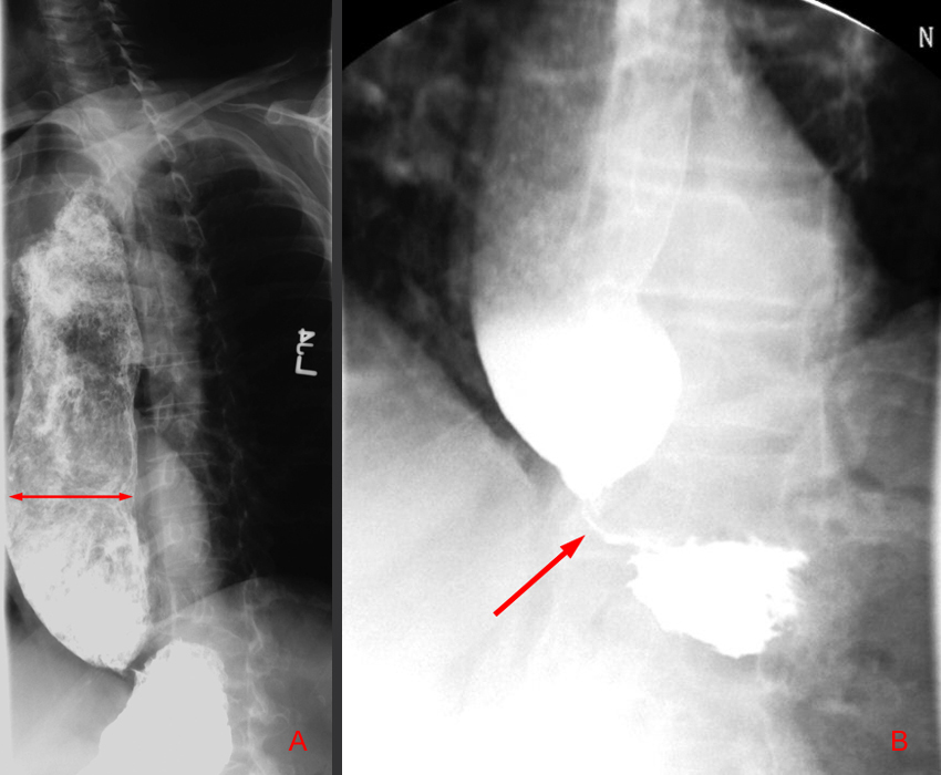
A 50-year-old gynecologist complains of dysphagia, regurgitation, and weight loss, and reports feeling as if food is stuck at the level of the xiphoid. An upright chest X-ray shows a dilated esophagus with an air-fluid level. Which of the following statements is false regarding achalasia?
Practice by Chapter
Esophageal Disorders
Practice Questions
Peptic Ulcer Disease
Practice Questions
Inflammatory Bowel Disease
Practice Questions
Irritable Bowel Syndrome
Practice Questions
Malabsorption Syndromes
Practice Questions
Pancreatitis (Acute and Chronic)
Practice Questions
Gastrointestinal Bleeding
Practice Questions
Liver Diseases and Cirrhosis
Practice Questions
Viral Hepatitis
Practice Questions
Biliary Tract Disorders
Practice Questions
Gastrointestinal Motility Disorders
Practice Questions
Gastrointestinal Malignancies
Practice Questions
Want unlimited practice?
Get full access to all questions, explanations, and performance tracking.
Scan to download app