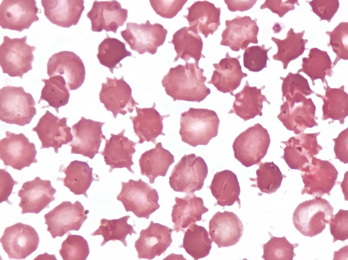
An adult male presented with a protruding abdomen, diarrhea, visual symptoms, and neurological manifestations. His LDL is low. Based on the peripheral smear finding shown in the image, what is the likely diagnosis?

A person is diagnosed with familial type IIa hyperlipoproteinemia. What is the basic defect in this type of hyperlipoproteinemia?
Which apolipoprotein is the primary structural component of LpA-I particles?
Which of the following enzymes is not targeted by hypolipidemic drugs?
What is the drug with the highest efficacy to increase plasma HDL?
Which of the following represents a desired lipid parameter for cardiovascular risk control in hypertension?
All of the following are true about nephrotic syndrome except?
HMG-CoA reductase is inhibited by:
Vitamin B12 deficiency can give rise to all of the following, except which of the following?
Which of these conditions is classified as a nephritic syndrome?
Explanation: ***Abetalipoproteinemia*** - The image shows **acanthocytes (spur cells)**, characterized by irregularly spaced, blunt projections, which are a hallmark of **abetalipoproteinemia** due to abnormal lipid metabolism and membrane defects. - The clinical presentation of a **protruding abdomen (steatorrhea/malabsorption)**, **diarrhea**, **visual symptoms (retinopathy)**, **neurological manifestations (ataxia, peripheral neuropathy)**, and **low LDL** all strongly point to abetalipoproteinemia, a disorder affecting the synthesis of B-apolipoprotein and chylomicrons. *EDTA changes* - **EDTA changes** typically manifest as **rouleaux formation**, platelet satellite formation, or cell shrinkage, with red blood cell morphology generally remaining normal in terms of spur cell formation. - These changes are **artifactual** and are not associated with the patient's systemic symptoms like malabsorption, neurological issues, or specific lipid profile findings. *Uremia* - While **uremia** can cause various red blood cell abnormalities, including **burr cells (echinocytes)** with regularly spaced, pointed projections, it generally does not cause the irregularly shaped **acanthocytes** seen in the image. - The systemic symptoms of uremia would primarily involve **renal dysfunction (e.g., elevated BUN, creatinine)**, which are not mentioned, and not specifically the **visual or malabsorption symptoms** seen here. *Burns* - Severe **burns** can lead to red blood cell fragmentation, causing **schistocytes** or **microspherocytes** due to heat-induced damage. - Burns are not typically associated with the formation of **acanthocytes** or the constellation of symptoms (malabsorption, neurological, visual) and lipid profile (low LDL) described in this patient. *Liver disease* - Advanced **liver disease (cirrhosis)** can cause **spur cell anemia** with acanthocytes due to altered cholesterol-to-phospholipid ratio in RBC membranes. - However, the key distinguishing feature is the **low LDL** in this patient, which is characteristic of abetalipoproteinemia, whereas liver disease typically does not present with specifically **low LDL** as a prominent feature. - Additionally, the constellation of **visual symptoms (retinopathy)** and **neurological manifestations** with malabsorption are more consistent with the fat-soluble vitamin deficiency (A, E, K) seen in abetalipoproteinemia rather than isolated liver pathology.
Explanation: ***Defective LDL receptor*** - **Familial hypercholesterolemia** (Type IIa hyperlipoproteinemia) is characterized by high levels of **LDL cholesterol** due to a genetic defect in the **LDL receptor** gene. - This defective receptor leads to impaired clearance of LDL particles from the bloodstream, resulting in their accumulation. *Lipoprotein lipase deficiency* - This defect is associated with **Type I hyperlipoproteinemia**, which is characterized by elevated **chylomicrons** and **triglycerides**, not primarily LDL cholesterol. - **Lipoprotein lipase (LPL)** is essential for the hydrolysis of triglycerides in chylomicrons and VLDL. *Abnormal activity of Apo E* - Variants of **Apolipoprotein E (Apo E)**, particularly Apo E2, are associated with **Type III hyperlipoproteinemia** (familial dysbetalipoproteinemia). - This condition involves increased levels of **chylomicron remnants** and **VLDL remnants** (IDL), not primarily isolated LDL elevation. *Overproduction of LDL* - While increased **LDL production** can contribute to elevated LDL levels, the primary genetic defect in familial type IIa hyperlipoproteinemia is strictly related to the impaired **clearance** of LDL due to a defective **LDL receptor**, rather than solely overproduction. - Many secondary causes of hypercholesterolemia can involve LDL overproduction, but Type IIa is specifically linked to the receptor defect.
Explanation: ***Apo A-I*** - **Apolipoprotein A-I (Apo A-I)** is the main structural and functional protein of **high-density lipoprotein (HDL)**. - It plays a crucial role in **reverse cholesterol transport**, facilitating the removal of excess cholesterol from peripheral tissues back to the liver. *Apo B-48* - **Apo B-48** is found exclusively in **chylomicrons**, which are responsible for transporting dietary lipids from the intestines. - It is synthesized in the **intestine** and is critical for the assembly and secretion of chylomicrons. *Apo A-II* - **Apo A-II** is another apolipoprotein found in HDL particles, but it is not the primary structural component. - While present, it is less abundant than Apo A-I and its precise role is still being researched, though it may influence **HDL metabolism**. *Apo B-100* - **Apo B-100** is the primary structural protein of **low-density lipoprotein (LDL)** and very-low-density lipoprotein (VLDL). - It is essential for the binding of LDL to the **LDL receptor**, mediating the uptake of cholesterol into cells.
Explanation: ***Peripheral decarboxylase*** - **Peripheral decarboxylase** (also known as DOPA decarboxylase) is involved in the synthesis of dopamine from L-DOPA and is a target for drugs used in **Parkinson's disease**, not hypolipidemic drugs. - Its inhibition by drugs like **carbidopa** or **benserazide** prevents the peripheral conversion of L-DOPA to dopamine, increasing L-DOPA availability for the brain. *HMG Co A reductase* - **HMG-CoA reductase** is the rate-limiting enzyme in cholesterol biosynthesis and is the primary target for **statins** (e.g., atorvastatin, simvastatin). - Statins effectively lower **LDL cholesterol** by inhibiting this enzyme, reducing endogenous cholesterol production. *Lipoprotein lipase* - **Lipoprotein lipase (LPL)** activity can be enhanced by certain hypolipidemic drugs, such as **fibrates**, which activate **PPAR-α**. - Increased LPL activity leads to enhanced hydrolysis of **triglycerides** from VLDL and chylomicrons, reducing triglyceride levels in plasma. *Acyl CoA, cholesterol acyl transferase 1* - **Acyl-CoA:cholesterol acyltransferase (ACAT) inhibitors** were developed as potential hypolipidemic agents to prevent cholesterol esterification and absorption. - While not widely used clinically due to efficacy and side effect profiles, **ACAT1** is involved in cholesterol esterification in the intestine and liver, making it a target for reducing cholesterol absorption.
Explanation: ***Nicotinic acid (Niacin)*** - Niacin has the **highest efficacy** among lipid-lowering drugs in increasing **plasma HDL cholesterol** levels, often by 15-35%. - It works by reducing the **hepatic synthesis of VLDL** (and thus LDL) as well as increasing the half-life of HDL. *Ezetimibe (Zetia)* - Ezetimibe primarily acts by inhibiting the **absorption of cholesterol** from the intestine. - While it lowers LDL cholesterol, its effect on **increasing HDL** is modest at best, typically in the single digits. *Gemfibrozil (Lopid)* - Gemfibrozil is a **fibrate** that is best known for significantly lowering **triglycerides** and increasing HDL cholesterol modestly. - Its effects on HDL are generally **less robust** than those of niacin, usually in the range of 10-20%. *Rosuvastatin (Crestor)* - Rosuvastatin is a **statin**, which primarily works by inhibiting **HMG-CoA reductase**, leading to a significant reduction in LDL cholesterol. - While statins can cause a small increase in HDL, typically about 5-10%, this effect is **not its primary mechanism** of benefit nor its greatest strength compared to niacin.
Explanation: Cholesterol/HDL < 3.5 [1] - A total cholesterol-to-HDL ratio of less than 3.5 is considered optimal for cardiovascular risk reduction. - This ratio indicates a favorable balance, where the proportion of 'good' HDL cholesterol is relatively high compared to total cholesterol. LDL / cholesterol > 10 mg% - This option is unclear and likely misphrased, as LDL cholesterol is typically measured independently, not as a ratio to total cholesterol in this manner [1]. - Desired LDL levels are typically much lower than 100 mg/dL for high-risk individuals, and a ratio of LDL to total cholesterol greater than 0.1 (or 10%) is generally observed, but not a specific target for reduction [1]. HDL < 30 mg% - An HDL level less than 40 mg/dL (or 30 mg% for some contexts) is considered low and undesirable, as high HDL is protective against cardiovascular disease [1]. - This value would indicate increased cardiovascular risk, contrary to a desired parameter. HDL / cholesterol ratio < 3.5 - This ratio, as stated, is the inverse of the commonly used and desirable total cholesterol-to-HDL ratio. - If the HDL/cholesterol ratio were less than 3.5, it would imply a relatively low HDL compared to total cholesterol, which is an undesirable cardiovascular risk factor [1].
Explanation: ***Decreased serum triglycerides*** - Nephrotic syndrome is characterized by **hyperlipidemia**, including **elevated total cholesterol** and **triglycerides**, due to increased hepatic synthesis of lipoproteins and decreased catabolism [1]. - This is a direct consequence of the body's attempt to compensate for low oncotic pressure and is a major diagnostic feature. *Hypoalbuminemia* - This is a **hallmark characteristic** of nephrotic syndrome, resulting from the significant loss of albumin in the urine [1]. - A low serum albumin level (typically <3.0 g/dL) contributes to **edema** due to decreased plasma oncotic pressure [1]. *Proteinuria >3.5 g/day* - This is the **defining diagnostic criterion** for nephrotic syndrome, indicating massive protein excretion through damaged glomerular capillaries [1]. - The protein loss is specifically defined as >3.5 grams per 1.73 m² of body surface area per day. *Increased risk of infection* - Patients with nephrotic syndrome are prone to infections, particularly **bacterial infections** like spontaneous bacterial peritonitis [1]. - This increased risk is due to the urinary loss of **immunoglobulins** (especially IgG), complement factors, and impaired cellular immunity [1].
Explanation: ***Lovastatin*** - **Lovastatin** is part of the statin class of drugs, which are potent competitive inhibitors of **HMG-CoA reductase**. - By inhibiting this enzyme, statins reduce the synthesis of **mevalonate**, a precursor to **cholesterol**, thereby lowering LDL-cholesterol levels. *Gemfibrozil* - **Gemfibrozil** is a **fibrate**, a class of drugs that primarily act by activating **peroxisome proliferator-activated receptor alpha (PPAR-α)**. - Its main effect is to decrease **triglyceride** levels and increase **HDL cholesterol**, not directly inhibit HMG-CoA reductase. *Clofibrate* - **Clofibrate** is also a **fibrate** and operates similarly to gemfibrozil by activating **PPAR-α**. - It primarily reduces **triglycerides** and has a modest effect on increasing HDL, but does not inhibit HMG-CoA reductase. *Nicotinic acid* - **Nicotinic acid** (niacin or vitamin B3) reduces hepatic synthesis of **VLDL** and inhibits the release of **fatty acids** from adipose tissue. - This leads to a decrease in **LDL cholesterol** and triglycerides, and an increase in **HDL cholesterol**, but it does not directly inhibit HMG-CoA reductase.
Explanation: ***Myopathy*** - **Myopathy**, or muscle disease, is not a direct consequence of **Vitamin B12 deficiency**. - **Vitamin B12 deficiency** primarily affects neurological and hematological systems due to its role in myelin synthesis and DNA production. [1] *Myelopathy* - **Myelopathy**, specifically subacute combined degeneration of the spinal cord, is a classic neurological complication of **Vitamin B12 deficiency**. - This involves demyelination of the posterior and lateral columns, leading to symptoms like **ataxia** and **sensory deficits**. *Optic atrophy* - **Optic atrophy** or **toxic amblyopia** can occur in severe cases of **Vitamin B12 deficiency**. - This damage to the **optic nerve** results in progressive vision loss. *Peripheral neuropathy* - **Peripheral neuropathy**, characterized by symptoms like **paresthesias**, numbness, and weakness, is a common neurological manifestation of **Vitamin B12 deficiency**. - It results from **demyelination** and axonal degeneration of peripheral nerves.
Explanation: ***Post infectious Glomerulonephritis*** - Characterized by **hematuria, hypertension, and edema**, typically following an infection, such as streptococcal pharyngitis [2]. - Immune-mediated response leads to **decreased GFR** and signs of nephritic syndrome [1][2]. *Focal segmental glomerulosclerosis* - Primarily causes **nephrotic syndrome**, characterized by proteinuria and edema rather than hematuria [2]. - Often associated with **secondary causes** like obesity or HIV, not typically post-infectious. *Membranous Glomerulopathy* - Results in significant **proteinuria** and is classified as a **nephrotic syndrome** rather than a nephritic one [2][3]. - It presents with **edema and hypoalbuminemia**, lacking the hallmark features of hematuria. *Minimal change disease* - Predominantly causes **nephrotic syndrome** with heavy proteinuria and little to no hematuria [2]. - Young children are commonly affected, and it responds well to **corticosteroid therapy** [1].
Get full access to all questions, explanations, and performance tracking.
Start For Free